
X-Ray Basics
X-rays are a form of electromagnetic radiation that can penetrate through the body, allowing for detailed imaging of internal structures. They were discovered by Wilhelm Conrad Roentgen in 1895 and have since become a vital tool in medical diagnostics. X-rays are a type of high-energy radiation that can pass through soft tissues but are absorbed by denser materials such as bones and metallic objects.
The basic principle behind x-ray imaging is the differential absorption of x-rays by different tissues. When an x-ray beam passes through the body, it is absorbed by the various tissues to varying degrees. The x-rays that pass through the body and reach the x-ray detector create an image that represents the differences in absorption. Dense structures, such as bones, appear white, while softer tissues appear in shades of gray, and air-filled spaces appear black.
This differential absorption allows healthcare professionals to visualize and diagnose conditions such as fractures, tumors, infections, and abnormalities in organs and tissues. X-rays are commonly used to examine the chest, bones, teeth, and certain internal organs.
It is important to note that x-rays are ionizing radiation, which means they can potentially cause cellular damage. However, the levels of radiation used in diagnostic x-ray imaging are carefully controlled to minimize risks and ensure patient safety. Protective measures, such as lead aprons and collimators, are used to shield sensitive areas from unnecessary radiation exposure.
In the next section, we will explore how a CT scanner utilizes x-rays to generate detailed cross-sectional images of the human body.
What is a CT Scanner?
A computed tomography (CT) scanner is a specialized medical imaging device that combines x-rays with advanced computer technology to produce detailed cross-sectional images of the body. CT scans provide a more comprehensive and three-dimensional view of the internal structures than traditional x-rays.
The CT scanner consists of a large, doughnut-shaped machine with a narrow table that can move patients through the opening. Inside the machine, an x-ray tube rotates around the patient, emitting a series of narrow beams that pass through the body from different angles. The beams are detected by an array of detectors positioned on the opposite side of the patient.
The CT scanner works by collecting a series of x-ray images as the rotating x-ray tube and detectors move around the patient. These images are then processed by a computer to create detailed cross-sectional slices of the body. These slices can be combined to generate three-dimensional images that provide a comprehensive view of the anatomy.
One of the major advantages of CT scanners is their ability to distinguish between different types of tissues based on their x-ray attenuations. This allows for enhanced visualization of blood vessels, organs, bones, and soft tissues. CT scans can help in the detection and diagnosis of various conditions, including cancers, injuries, infections, and abnormalities.
Over the years, CT technology has advanced significantly, with the development of spiral CT and multidetector CT systems. Spiral CT enables continuous acquisition of x-ray images while the patient is moving through the machine, resulting in faster scan times. Multidetector CT systems utilize multiple rows of detectors, allowing for even faster scanning and higher resolution images.
How Does X-Ray Imaging Work?
X-ray imaging is a widely used medical diagnostic technique that allows healthcare professionals to visualize and assess the internal structures of the body. Understanding how x-ray imaging works can help us appreciate the process of capturing detailed images using x-rays.
The basic principle behind x-ray imaging is the differential absorption of x-rays by various tissues in the body. When an x-ray beam is directed towards the body, it passes through the tissues before reaching a detector on the other side. As the x-rays pass through the body, they interact with the tissues and are absorbed to different degrees depending on the density of the tissue.
Dense tissues, such as bones, absorb more x-rays and appear white on the resulting image. Softer tissues, like muscles and organs, absorb fewer x-rays and appear as shades of gray. Air-filled spaces, like the lungs, allow most of the x-rays to pass through, resulting in dark areas on the image.
To capture an x-ray image, the patient is positioned between the x-ray source and the detector. The x-ray machine emits a focused beam of x-rays that passes through the patient’s body, creating a shadow of the internal structures on the detector. The detector converts the x-rays into an electrical signal, which is then processed by a computer to create a visible image.
Factors such as the patient’s size, thickness, and density of the tissues, as well as the energy and intensity of the x-ray beam, affect the quality and clarity of the image. The positioning of the patient during the x-ray examination is also crucial to obtain the desired view of the targeted area.
It’s important to note that x-ray imaging involves minimal discomfort and is generally considered safe when performed in controlled and appropriate settings. However, precautions are taken to minimize radiation exposure, especially for pregnant women and children.
In the next section, we will explore the technology behind CT scanning, which utilizes x-ray images to create detailed cross-sectional images of the body.
The Technology Behind CT Scanning
Computed tomography (CT) scanning is a sophisticated medical imaging technology that combines x-rays and advanced computer processing to create detailed cross-sectional images of the body. This section will delve into the intricate technology that makes CT scanning possible.
At the heart of a CT scanner is a rotating x-ray tube, which emits narrow beams of x-rays as it spins around the patient. These x-rays pass through the patient’s body and are detected by an array of detectors positioned on the opposite side. The detectors measure the intensity of the x-rays that pass through the body at various angles.
The x-ray tube and detectors are synchronized with the computer to acquire multiple x-ray images as the scanner rotates. These images capture the attenuation of the x-rays by different tissues in the body, varying from highly attenuating structures like bones to less attenuating soft tissues and air-filled spaces.
Once the x-ray images are acquired, the computer uses a process called image reconstruction to transform the acquired data into detailed cross-sectional images of the body. This reconstruction involves complex algorithms that calculate the relative attenuation values and spatial relationships of each pixel in the acquired images.
Modern CT scanners use a technique called spiral or helical scanning, where the x-ray tube and detectors continuously move together in a helical path along the length of the body. This continuous motion allows for the acquisition of a seamless volume of image data, which can be used to generate three-dimensional images and perform advanced imaging techniques.
The rapid advancement of technology has led to the development of multidetector CT scanners, which feature multiple rows of detectors. These scanners can acquire multiple slices of data simultaneously, resulting in faster scan times and improved image quality. Additionally, advancements in computer processing power have enabled faster reconstruction times and more advanced image manipulation capabilities.
CT scanning technology continues to evolve, with ongoing research and development focused on improving image quality, reducing radiation exposure, and expanding the clinical applications. The advancements in CT scanning technology have revolutionized medical diagnostics and significantly contributed to the accurate diagnosis and treatment of various medical conditions.
The Role of X-Ray Tubes
X-ray tubes play a crucial role in computed tomography (CT) scanning, serving as the source of the x-ray beams used to generate detailed images of the body. This section will explore the importance and functionality of x-ray tubes in CT imaging.
An x-ray tube is a specialized electronic device that converts electrical energy into x-ray radiation. It consists of a cathode and an anode enclosed in a vacuum-sealed glass or metal enclosure. The cathode emits a beam of electrons, which are accelerated towards the anode by a high-voltage potential.
When the high-speed electrons from the cathode interact with the anode, they suddenly decelerate, releasing energy in the form of x-ray photons. These photons form the x-ray beam that passes through the patient’s body during a CT scan.
X-ray tubes used in CT scanners are designed to produce high-intensity and collimated x-ray beams. High-intensity beams ensure that enough x-rays reach the detectors to generate clear and detailed images. Collimation refers to the process of narrowing the x-ray beam to a specific area, allowing for precise imaging of targeted regions and reducing unnecessary radiation exposure to surrounding tissues.
One important aspect of x-ray tubes in CT scanning is their ability to generate x-ray beams of different energies or kilovoltages (kVp). The choice of kilovoltage depends on the type of examination and the density of the tissues being imaged. Higher kilovoltages are suitable for examining denser tissues, such as bones, while lower kilovoltages are used for imaging softer tissues.
Additionally, x-ray tubes in modern CT scanners feature the ability to rapidly switch on and off, enabling the acquisition of multiple x-ray projections at various angles. This projection data is then used by the computer to reconstruct detailed cross-sectional images of the body.
X-ray tubes in CT scanners must also be able to withstand the high heat generated during operation. Heat management is critical to preventing overheating and maintaining the optimal functioning of the tube. Special cooling systems, such as oil or water, are used to dissipate the heat generated by the tube during prolonged use.
Advancements in x-ray tube technology have led to increased efficiency, faster scan times, and improved image quality in CT scanning. Ongoing research and development aim to further enhance the performance and longevity of x-ray tubes, contributing to more accurate diagnosis and better patient care.
Detectors and their Importance
Detectors play a vital role in computed tomography (CT) scanning by capturing the x-rays that pass through the body and converting them into electrical signals. This section will delve into the importance of detectors in CT imaging and their role in producing detailed and accurate images.
In CT scanners, detectors are positioned opposite the x-ray tube, aligned to receive the x-rays that pass through the patient’s body. These detectors consist of arrays of scintillation crystals or semiconductor detector materials that absorb the x-rays and convert them into electrical signals.
One of the primary functions of detectors in CT scanning is to accurately measure the intensity of the transmitted x-rays at various angles. These measurements are crucial in reconstructing cross-sectional images of the body. The detectors capture the attenuated x-rays that have passed through the body, providing information about the different densities of tissues.
Detectors in modern CT scanners are capable of detecting even the slightest variations in x-ray intensity, resulting in high-resolution images. They play a vital role in producing detailed images with a wide dynamic range, allowing for the detection of subtle differences between tissues.
The speed and efficiency of the detectors are also crucial in CT scanning. Rapid data acquisition is required to capture multiple projections as the x-ray tube rotates around the patient. High-speed detectors ensure that the necessary information is collected in a timely manner, reducing the time required for image acquisition.
The ability of detectors to measure the intensity of x-rays accurately is crucial for dose optimization. With precise measurements, CT scanners can be equipped with automatic exposure control mechanisms that adjust the x-ray output based on the patient’s size, thickness, and other factors. This helps minimize radiation exposure while maintaining optimal image quality.
The advancements in detector technology have led to the development of multidetector CT scanners. These scanners feature multiple rows or layers of detectors, allowing for faster acquisition of image data. With more detectors, larger volumes of data can be acquired simultaneously, resulting in shorter scan times and improved spatial resolution.
Overall, detectors are indispensable components of CT scanners, enabling the conversion of x-rays into electrical signals and providing essential information for image reconstruction. Their ability to accurately measure x-ray intensity and capture subtle variations in tissue density contributes to the generation of detailed and precise diagnostic images.
The Process of CT Scanning
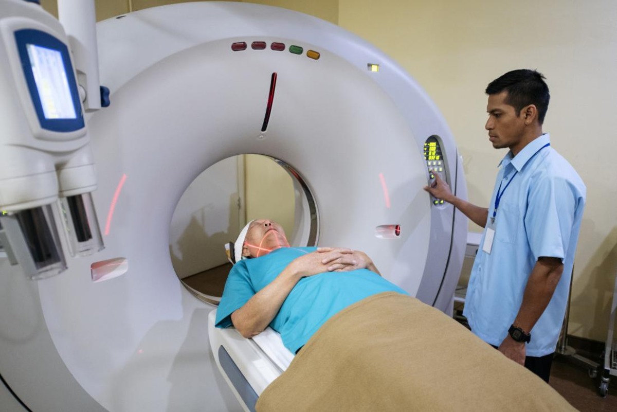
The process of CT scanning involves multiple steps, including patient preparation, positioning, data acquisition, and image reconstruction. This section will outline the key stages in a typical CT scan, providing an overview of how the imaging process unfolds.
Before the scan, the patient may be required to change into a hospital gown and remove any metallic objects or jewelry that may interfere with the scan. Depending on the type of examination, the patient may also need to fast or drink contrast agents to enhance the visibility of specific structures.
Once prepared, the patient lies down on the CT scanning table, which is then positioned to ensure the targeted area is properly aligned with the x-ray beam. The technologist ensures the patient’s comfort and provides instructions on breath-holding or remaining still during the scan to avoid motion artifacts.
When the scan begins, the CT scanner’s rotating x-ray tube and array of detectors revolve around the patient, capturing multiple x-ray projections from different angles. The x-ray machine emits narrow beams of x-rays, which pass through the body and are detected on the opposite side. The detectors collect the attenuation data and send it to the computer for further processing.
During the scan, the patient may hear humming or clicking sounds as the x-ray tube and detectors move. Although the process is painless, the patient may be asked to hold their breath for a brief moment to minimize motion artifacts, particularly when capturing images of the chest or abdomen.
Once the data acquisition is complete, the computer processes the collected information to reconstruct cross-sectional images. This process involves intricate algorithms that calculate the relative attenuation of the x-rays as they passed through the body at different angles. The computer then generates images that depict the varying densities of the tissues.
After the image reconstruction, the radiologist or a healthcare professional reviews the images to make a diagnosis. These images provide detailed information about the internal structures, allowing the healthcare team to assess and detect abnormalities or conditions. The images can be viewed in various planes, such as axial, sagittal, or coronal, providing a comprehensive view of the anatomy.
The entire CT scanning process typically lasts a few minutes, depending on the targeted area and the complexity of the examination. Once the scan is complete, the patient can usually resume their normal activities without any restrictions or side effects.
Advancements in CT scanning technology, such as multidetector scanners and faster reconstruction algorithms, have significantly reduced scanning times while maintaining excellent image quality. The process has become more efficient, offering healthcare professionals a valuable tool for accurate diagnosis and treatment planning.
The Image Reconstruction Process
The image reconstruction process is a critical step in computed tomography (CT) scanning that converts the acquired raw data into detailed and meaningful cross-sectional images of the body. This section will explain the key aspects of the image reconstruction process in CT scanning, highlighting its importance in producing accurate diagnostic images.
When a CT scan is performed, the x-ray machine rotates around the patient, capturing a series of x-ray projections from various angles. These projections provide valuable data about how the x-rays were attenuated as they passed through the different tissues in the body.
The raw data collected by the detectors is a vast amount of attenuation values measured at numerous points across the patient’s body. To transform this data into meaningful images, a process called image reconstruction is employed.
The image reconstruction process involves complex mathematical algorithms that analyze the raw data and generate high-quality images. The software uses these algorithms to calculate the relative attenuation values for each pixel in the image based on the measured x-ray intensities and the known properties of the tissues being imaged.
The reconstruction algorithms take into account factors such as the position of the detectors, the geometry of the scan, and the physical properties of the x-ray beam. These algorithms employ advanced mathematical techniques, such as filtered back projection or iterative reconstruction, to accurately map the attenuation values and create detailed cross-sectional images.
Filtered back projection is the most commonly used reconstruction technique in CT scanning. It involves passing the raw data through various filters to remove unwanted noise and artifacts, and then back projecting the filtered data to generate the final image. This process iterates multiple times to refine the image quality.
Iterative reconstruction, on the other hand, utilizes mathematical models and iterations to refine the image reconstruction process. It iteratively matches the simulated projections to the measured projections to reconstruct a highly accurate image.
The image reconstruction process allows for the visualization of various tissues within the body. Each pixel in the resulting images represents a specific attenuation value, which corresponds to the density of the tissue at that particular location.
After the image reconstruction is complete, the radiologist or a healthcare professional can analyze the cross-sectional images to identify any abnormalities or conditions that require further evaluation. The images can be viewed and manipulated in different planes, providing comprehensive views of the anatomy.
Advancements in computational power and reconstruction algorithms have significantly improved the speed and accuracy of the image reconstruction process. The advancements have also led to the development of advanced imaging techniques, such as dual-energy CT and dynamic imaging, which further enhance the diagnostic capabilities of CT scanning.
Advantages of CT Scanning
Computed Tomography (CT) scanning offers several advantages in medical imaging, making it a valuable tool for diagnostic and treatment purposes. This section will outline the key advantages of CT scanning and how it contributes to improved patient care.
One of the significant advantages of CT scanning is its ability to provide detailed and highly accurate cross-sectional images of the body. CT scans produce images with excellent spatial resolution, allowing healthcare professionals to visualize and assess anatomical structures with greater precision compared to other imaging techniques.
CT scans are versatile and can be used to examine various parts of the body, including the head, chest, abdomen, pelvis, and musculoskeletal system. This wide range of applications makes CT scanning a valuable diagnostic tool for detecting and diagnosing a variety of conditions.
The speed of CT scans is another advantage, with modern CT scanners capable of capturing images in a matter of seconds. This rapid acquisition time is particularly beneficial for patients who may have difficulty remaining still or holding their breath for an extended period, such as pediatric or critically ill patients.
CT scanners can generate images in multiple planes, including axial, sagittal, and coronal. These planes provide healthcare professionals with a comprehensive view of the anatomy, ensuring a more accurate assessment of the patient’s condition and assisting in treatment planning.
One of the significant advantages of CT scanning is its ability to distinguish between different tissues based on their densities. This allows for enhanced visualization of bones, blood vessels, organs, and soft tissues. With CT scans, healthcare professionals can detect and characterize various conditions, such as tumors, fractures, infections, and abnormalities.
With the introduction of multidetector CT scanners, the time required for acquiring images has significantly reduced. These scanners can capture multiple slices of data simultaneously, resulting in shorter scan times. This faster data acquisition is particularly advantageous for patients who may have limited tolerability for longer examinations.
CT scans can provide valuable information for guiding interventions and treatments. CT-guided procedures, such as biopsies or drainages, allow for precise needle placement under real-time imaging guidance, minimizing risks and improving outcomes. CT scans can also be used for radiation therapy planning, assisting in the accurate targeting of tumors while sparing healthy surrounding tissues.
Additionally, advancements in CT scanning technology have led to the development of functional imaging techniques, such as perfusion CT and CT angiography. These techniques provide valuable information about blood flow, tissue viability, and vascular structures, enhancing the ability to evaluate conditions such as stroke, tumors, and vascular diseases.
Overall, CT scanning offers numerous advantages in terms of its ability to produce detailed images, its versatility in imaging different anatomical areas, its rapid acquisition time, and its usefulness in guiding interventions and treatments. These advancements in CT scanning technology have revolutionized medical diagnostics and significantly contributed to improved patient care.
Limitations and Risks of CT Scanning
While computed tomography (CT) scanning is a valuable imaging tool, it is essential to be aware of its limitations and associated risks. This section will discuss the potential drawbacks and considerations of CT scanning in medical diagnostics and patient care.
One limitation of CT scanning is its exposure to ionizing radiation. CT scans utilize x-rays, which are a form of ionizing radiation that can potentially cause cellular damage. Although the radiation doses in CT scans are carefully controlled and kept as low as reasonably achievable, repeated or unnecessary exposure may increase the risk of radiation-related effects, such as an increased likelihood of developing cancer later in life.
The risk associated with radiation exposure is higher in certain groups, such as pregnant women and children, as they are more sensitive to radiation. Special considerations must be taken to minimize radiation exposure in these populations, such as using alternative imaging modalities or adjusting scanning parameters to reduce radiation dose without compromising image quality.
Another limitation of CT scanning is the potential for false-positive or false-negative results. CT scans provide detailed images, but interpretation errors can occur, leading to incorrect or inconclusive diagnoses. In some cases, additional diagnostic tests or procedures may be necessary to confirm or refine the findings observed on CT scans.
CT scanning involves the use of contrast agents to enhance the visualization of certain structures or abnormalities. While generally safe, some individuals may experience an allergic reaction to these contrast agents. Patients with known allergies to iodine-based contrast agents or a history of adverse reactions should inform their healthcare provider prior to the CT scan.
Special considerations should also be taken for patients with impaired kidney function, as contrast agents can potentially cause kidney damage. Precautions, such as hydration and the use of alternative contrast agents, may be necessary for patients at risk of renal complications.
In certain cases, CT scanning may not be suitable for patients with claustrophobia or who are unable to remain still during the examination. Open-air or wide-bore CT scanners may be options worth considering to accommodate these patients while still obtaining the necessary diagnostic information.
Finally, cost and availability may be considered as limitations, as the equipment and expertise required for CT scanning can be relatively expensive and may not be accessible in all healthcare settings. This may result in limited availability or longer wait times for patients needing CT scans.
It is important for healthcare professionals to weigh the benefits against the potential risks of CT scanning for each individual patient. Alternative imaging modalities may be considered when appropriate to minimize radiation exposure or address specific limitations or contraindications.
Despite these limitations and risks, CT scanning remains a valuable tool in medical diagnostics, providing detailed and accurate images in many clinical scenarios. Ongoing research and advancements in technology aim to further reduce radiation dose, enhance image quality, and improve patient safety in CT scanning.