
Magnetic Resonance Imaging (MRI)
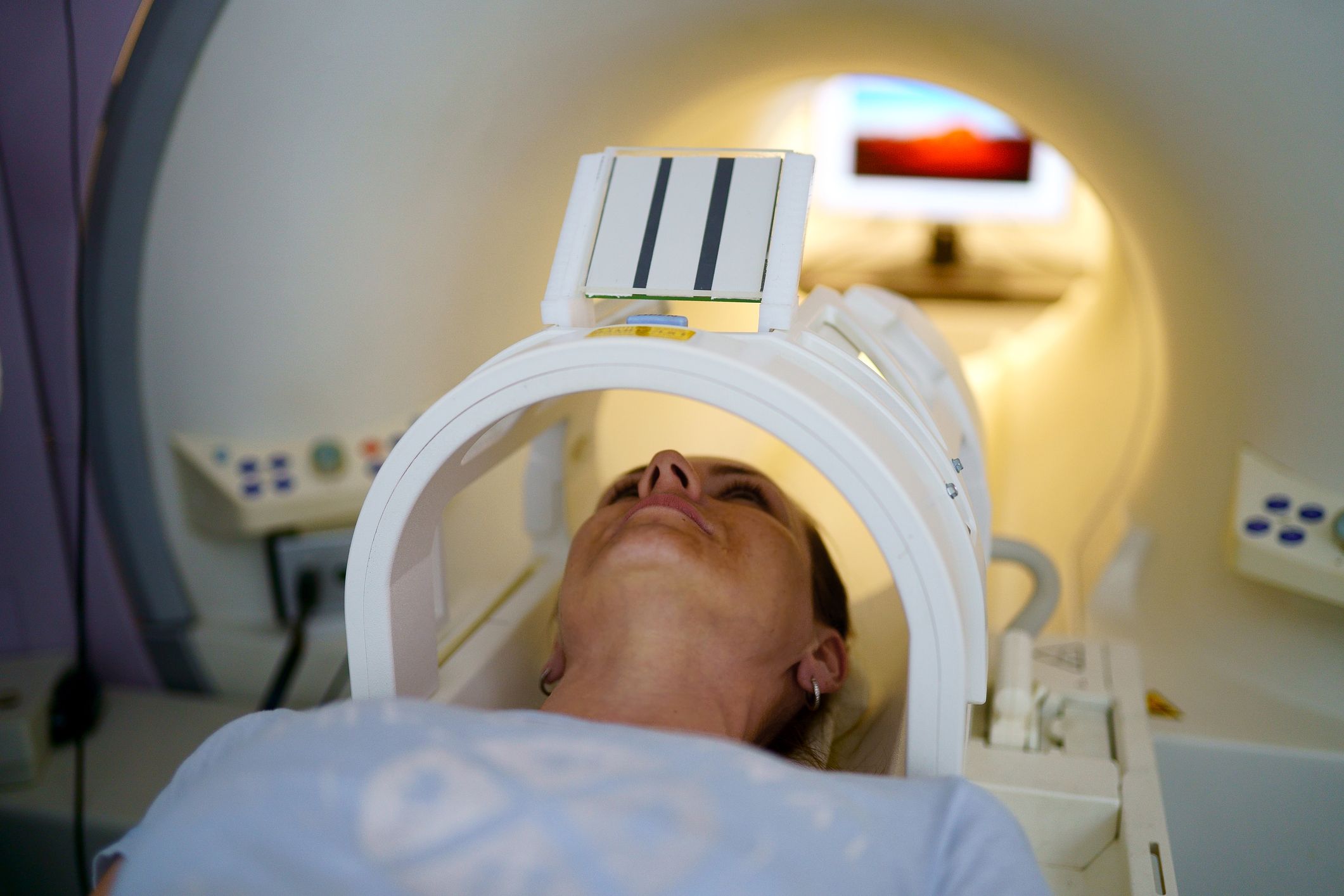
Magnetic Resonance Imaging (MRI) is a non-invasive medical imaging technique that provides detailed pictures of the internal structures of the body. It uses a combination of strong magnetic fields and radio waves to generate high-resolution images, allowing healthcare professionals to visualize soft tissues, organs, and bones with exceptional clarity.
Unlike other imaging techniques such as X-rays or CT scans, which use ionizing radiation, MRI is considered safer as it does not expose patients to harmful radiation. Instead, it utilizes the properties of hydrogen atoms in our bodies.
At the heart of MRI technology is the phenomenon of nuclear magnetic resonance (NMR). When a patient is placed inside the MRI scanner, the magnetic fields cause the hydrogen atoms in the body to align either with or against the applied field. This alignment creates a net magnetization that can be manipulated by the scanner to produce a measurable signal.
The scanner uses powerful magnets to create a strong and uniform magnetic field. This main magnetic field is produced by superconducting coils that are cooled to extremely low temperatures to eliminate any electrical resistance. The strength of the field is measured in teslas (T), with higher field strengths providing higher image resolution.
Additionally, the MRI system incorporates gradient coils, which produce smaller, varying magnetic fields. These gradients help to spatially localize the signals from different regions of the body. By applying varying magnetic gradients, the scanner can precisely determine the location of the signals and construct detailed images.
Basics of Magnetic Resonance
To understand how MRI works, it’s essential to grasp the fundamentals of magnetic resonance. In an MRI scanner, the aligning and realignment of hydrogen atoms play a crucial role in generating the signal used to create the images.
Hydrogen atoms, which are abundant in the human body due to the high water content, consist of a single proton. The protons possess an intrinsic property known as spin, which gives them a tiny magnetic moment. In the absence of an external magnetic field, the spins of the protons are randomly oriented.
When a patient is placed inside the MRI scanner, a powerful magnetic field is applied to their body. This external magnetic field causes the spins of the hydrogen atoms to align either parallel or anti-parallel to the field. The majority of the spins align parallel, resulting in a net magnetization vector along the direction of the external field.
A radiofrequency pulse is then directed towards the patient. This pulse delivers a specific amount of energy to the hydrogen atoms, causing them to absorb the energy and temporarily tip their spins away from the alignment with the external field. The frequency of the pulse corresponds to the resonance frequency of the hydrogen protons in the magnetic field.
After the radiofrequency pulse, the hydrogen spins gradually return to their original alignment within the magnetic field. As they do so, they release the absorbed energy in the form of a radiofrequency signal. This signal is detected by receiver coils positioned around the patient’s body.
The detected signal is then processed to create an image. By manipulating the timing and characteristics of the radiofrequency pulses, different contrasts and image types can be produced. Tissue properties such as relaxation times and proton density influence the signal intensity in the resulting images.
The Role of Magnets in MRI
Magnets are at the core of magnetic resonance imaging (MRI) technology. They play a critical role in creating a strong and uniform magnetic field that is essential for accurate imaging of the human body.
In an MRI system, there are typically one or more superconducting magnets. These magnets are cooled to extremely low temperatures using liquid helium in order to achieve superconductivity, which eliminates electrical resistance. The superconducting magnets are housed within a cylindrical structure, forming the bore of the MRI scanner.
The main magnet is responsible for generating a strong and uniform magnetic field. The strength of this magnetic field is measured in teslas (T), with higher field strengths offering better image quality and resolution. Most clinical MRI scanners operate at field strengths of 1.5 Tesla or 3 Tesla.
The main magnetic field aligns the proton spins of the hydrogen atoms in the patient’s body. These protons possess an intrinsic magnetic moment and tend to align either parallel or anti-parallel to the magnetic field. The majority of protons align parallel, creating a net magnetization vector.
In addition to the main magnet, gradient coils are employed in MRI systems. These gradient coils produce smaller, varying magnetic fields in different directions. They are placed within the main magnetic field and allow for precise spatial localization of the signals emitted by the hydrogen atoms.
By applying gradient fields, the MRI scanner can encode spatial information into the detected signals. This information is crucial in reconstructing detailed images of the internal structures of the body. Different gradient strengths and directions are used to obtain images in various planes, such as axial, sagittal, and coronal.
Furthermore, the magnets in the MRI system require careful calibration and maintenance to ensure the generation of a stable and homogeneous magnetic field. Any fluctuations or inhomogeneities in the magnetic field can lead to image artifacts and affect the accuracy of the diagnostic information.
Overall, magnets are the backbone of MRI technology, providing the necessary magnetic field to align the proton spins and enable the acquisition of detailed images. The advancements in magnet technology have contributed significantly to the improvement of MRI image quality and the diagnostic capabilities of this valuable medical imaging modality.
How Gradient Coils are Utilized
Gradient coils are an integral part of magnetic resonance imaging (MRI) systems, working in tandem with the main magnet to provide precise spatial information. These coils produce varying magnetic fields that are essential for encoding spatial details into the detected signals.
In an MRI scanner, gradient coils are typically placed within the main magnetic field, surrounding the patient. They consist of multiple sets of coils: X, Y, and Z, corresponding to the three-dimensional axes.
The gradient coils are responsible for creating magnetic field gradients in different directions. These gradients allow the scanner to precisely localize the origin of the emitted magnetic signals, enabling the reconstruction of detailed images in various planes.
By applying different currents to the gradient coils, the strength and direction of the magnetic field gradients can be controlled. For example, if a higher current is applied to the X-axis gradient coil, a stronger magnetic field gradient in the X direction is produced.
During the imaging process, the gradient coils are activated in a carefully timed manner. When the hydrogen atoms in the patient’s body release their absorbed energy after the radiofrequency excitation, the gradient coils create additional magnetic field gradients that vary in strength and direction.
These varying gradients cause the hydrogen atoms at different locations within the body to resonate at different frequencies. By encoding this information into the detected signals, the scanner can precisely determine the spatial position of each signal and construct detailed images.
The gradient coils are particularly crucial for obtaining images in different planes. For example, by utilizing the X-axis and Y-axis gradients, the scanner can acquire axial images that show cross-sections of the body. By using the Z-axis gradient, images in the sagittal or coronal planes can be obtained.
It’s essential to carefully calibrate and maintain the gradient coils to ensure their accuracy and stability. Any irregularities or fluctuations in the gradients can result in imaging artifacts and compromise the image quality.
Overall, the gradient coils in MRI systems play a vital role in providing precise spatial information, allowing for detailed and accurate imaging of the internal structures of the body. Without these coils, the MRI scanner would be unable to reconstruct the spatial information necessary for producing high-quality images.
Radiofrequency Coils and their Function
Radiofrequency (RF) coils are essential components of magnetic resonance imaging (MRI) systems, working in conjunction with the main magnet and gradient coils to acquire the radiofrequency signals emitted by the patient’s body. These coils play a crucial role in capturing the signals and facilitating the creation of high-resolution images.
In an MRI scanner, RF coils are positioned around the area of the body being imaged. They are designed to transmit the radiofrequency pulses that excite the hydrogen atoms in the patient’s tissues and receive the resulting signals emitted during relaxation.
Transmit RF coils are responsible for delivering the radiofrequency pulses to the targeted area. These coils generate a uniform and homogenous radiofrequency field that is needed to excite the hydrogen atoms for imaging. The transmitted pulses are carefully timed and controlled to achieve the desired imaging parameters.
Receive RF coils, on the other hand, are used to capture the emitted radiofrequency signals from the patient’s tissues. These signals encode information about the relaxation times and proton density of the tissues and are crucial for image reconstruction.
RF coils can vary in design and configuration depending on the specific application and body part being imaged. Coils may range from surface coils that are placed directly on the body surface to internal coils that are inserted into body cavities or positioned near targeted structures.
The design and placement of RF coils are critical factors in achieving good signal-to-noise ratio (SNR) and image quality. SNR refers to the strength of the received signal compared to the background noise. Coils are carefully positioned to optimize the SNR, ensuring better image resolution and diagnostic accuracy.
In some cases, multiple RF coils may be used for simultaneous image acquisition. This technique, known as parallel imaging, enhances the imaging speed and reduces scan time. By employing arrays of RF coils with different sensitivities and spatial coverage, parallel imaging can provide fast and high-resolution imaging of larger areas.
It’s worth noting that RF coils need to be carefully selected and matched to the specific clinical application. Factors such as coil size, coil sensitivity, and coil shape can impact the image quality and diagnostic accuracy. The continual advancements in RF coil technology have greatly contributed to improving the overall imaging capabilities of MRI systems.
The Process of Image Acquisition
The process of image acquisition in magnetic resonance imaging (MRI) involves several steps that work together to capture the signals emitted by the patient’s body and convert them into detailed images. This process includes signal generation, signal detection, and image reconstruction.
When a patient is placed inside the MRI scanner, the main magnet and gradient coils are activated to create a strong and uniform magnetic field. This magnetization aligns the hydrogen atoms within the patient’s body, setting the stage for signal generation.
The next step is the excitation of the hydrogen atoms using a radiofrequency (RF) pulse. The RF pulse, transmitted through the RF coil, energizes the hydrogen atoms and causes them to temporarily deviate from their equilibrium state. This excitation process is carefully timed and tailored to achieve the desired imaging parameters.
After the excitation, the hydrogen atoms begin to relax and return to their equilibrium state. As they relax, they emit radiofrequency signals that contain information about the tissue properties and spatial distribution. These emitted signals are captured by the RF receive coil, which acts as a sensitive detector.
The detected signals are weak and require amplification and filtering to enhance their strength and quality. Once amplified and filtered, the signals are digitized, converting them from analog to digital form. This digital data is then stored for further processing and image reconstruction.
Image reconstruction is a crucial step in the process of image acquisition. The digitized data from the detector coils is processed using advanced mathematical algorithms on a computer. These algorithms analyze the acquired signals, correct for various artifacts, and reconstruct the data into a series of cross-sectional images.
During the image reconstruction process, additional processing techniques can be applied to enhance the quality and diagnostic value of the images. These techniques include noise reduction, contrast adjustments, and three-dimensional rendering.
The final result of the image acquisition process is a set of high-resolution images that provide a detailed depiction of the internal structures of the body. These images can be used to aid in the diagnosis and monitoring of various medical conditions.
It’s important to note that the entire image acquisition process is carried out in a matter of seconds or minutes, depending on the specific imaging protocol. The speed and efficiency of MRI systems have contributed to their widespread use in medical imaging.
Pulse Sequences and their Importance
Pulse sequences are fundamental components of magnetic resonance imaging (MRI) that determine the timing and characteristics of the radiofrequency (RF) pulses and magnetic gradients used to acquire the signals and create the image. These sequences play a crucial role in shaping the contrast, resolution, and diagnostic capabilities of MRI scans.
There are various types of pulse sequences used in MRI, each designed to emphasize different tissue properties or highlight specific anatomical details. The selection of an appropriate pulse sequence depends on the clinical question and the desired imaging goals.
One commonly used pulse sequence is the spin-echo sequence. It involves a 90-degree RF pulse followed by a 180-degree RF pulse, with a delay in between known as the echo time (TE). The spin-echo sequence allows for the measurement of T1 and T2 relaxation times, which provide valuable information about tissue characteristics and pathology.
Another important pulse sequence is the gradient-echo sequence. In this sequence, the RF pulse is applied in the presence of a magnetic gradient, resulting in the acquisition of gradient-echo images. These images have different contrast properties compared to spin-echo images and are particularly useful for capturing dynamic processes, such as blood flow and perfusion imaging.
Furthermore, advanced pulse sequences like the fast spin-echo sequence (FSE) and echo-planar imaging (EPI) have revolutionized MRI imaging by significantly reducing scan times. FSE allows for rapid acquisition of multiple echoes, resulting in shorter scan durations, while EPI enables imaging of the entire body in a fraction of the time compared to traditional methods.
Pulse sequences also play a crucial role in improving the quality and diagnostic value of MR images. Techniques such as fat suppression, diffusion-weighted imaging, and contrast-enhanced imaging can be incorporated into the pulse sequences to enhance the visualization of certain tissues or highlight abnormalities.
Moreover, pulse sequences are essential for determining the image resolution and spatial information. By manipulating the timing and amplitude of the magnetic gradients, the MRI scanner can acquire images in different orientations (axial, sagittal, or coronal) and at various slice thicknesses, catering to the specific clinical need.
Overall, pulse sequences are vital in MRI as they dictate the timing, contrast, resolution, and diagnostic capabilities of the scans. The selection of an appropriate pulse sequence is tailored to the specific clinical question, optimizing image acquisition and providing crucial information for accurate diagnosis and treatment planning.
The Role of a Computer in Image Reconstruction
A computer plays a crucial role in the process of image reconstruction in magnetic resonance imaging (MRI). Once the raw data is acquired from the scanner, it is processed and transformed into detailed images through complex algorithms and computations.
The first step in the image reconstruction process is the digitization of the acquired data. The analog signals captured by the receive coils are converted into digital format using an analog-to-digital converter. This digital data is then fed into the computer for further processing.
The raw data received from the scanner is in the form of a series of complex numbers known as k-space data. Each complex number represents the signal strength and phase information from a specific location in the body. The computer performs Fourier transforms and other mathematical operations on this data to convert it into the spatial domain.
The computer uses advanced algorithms to process the k-space data and reconstruct the image. One of the commonly used algorithms is the Fourier transform, which converts the frequency-domain information into the spatial-domain information. Other reconstruction algorithms, such as filtered back projection and iterative reconstruction, are also used to enhance image quality and reduce artifacts.
During the reconstruction process, the computer takes into account various factors such as the pulse sequence, gradient strengths, coil sensitivities, and other acquisition parameters. These factors are utilized to correct for distortions, artifacts, and signal variations that may occur during the imaging process.
The computer also applies imaging parameters such as slice thickness, field of view, and image resolution to generate images that meet the specific clinical requirements. Sophisticated reconstruction techniques can further improve image quality by reducing noise, increasing contrast, and enhancing spatial resolution.
Once the images are reconstructed, the computer allows for further manipulation and analysis. Radiologists can use specialized software tools to adjust image settings, perform measurements, and apply post-processing techniques for better visualization and interpretation. The computer also enables the storage and retrieval of the reconstructed images for future reference.
With the advancements in computing power and software algorithms, image reconstruction in MRI has become faster and more accurate, leading to improved diagnostic capabilities. The ability of the computer to process and reconstruct vast amounts of data in real-time has revolutionized the field of medical imaging and greatly benefited patient care.
Challenges and Limitations of MRI
Magnetic Resonance Imaging (MRI) is a powerful medical imaging technique that offers many advantages over other imaging modalities. However, it also has its own set of challenges and limitations that need to be considered in clinical practice.
One significant challenge with MRI is its high cost. The complex machinery, including the superconducting magnets and sophisticated computer systems, along with the need for specialized infrastructure, contribute to the high upfront and maintenance costs of MRI scanners. These costs can limit accessibility to MRI technology in certain healthcare facilities.
MRI examinations can also be time-consuming. The process of image acquisition takes longer compared to other imaging modalities such as X-rays or CT scans. Multiple sequences and adjustments may be required to capture the desired images, resulting in longer examination times. This can be challenging for patients who may experience discomfort, claustrophobia, or difficulty lying still for an extended period.
MRI scanning is contraindicated for individuals with certain conditions or devices. Patients with ferromagnetic implants, such as pacemakers or cochlear implants, may not be able to undergo MRI due to safety concerns. Additionally, patients with claustrophobia or anxiety may find it challenging to tolerate the confined space of the MRI scanner, potentially requiring the use of sedation or alternative imaging methods.
Motion artifacts are another limitation of MRI. Even slight movement of the patient during the imaging process can result in blurred or distorted images. This can be particularly problematic when imaging dynamic structures or patients who are unable to remain still for an extended period.
Furthermore, MRI is not suitable for all types of imaging. It may not be the ideal modality for evaluating certain conditions, such as acute trauma or bony structures, where other imaging techniques like X-rays or CT scans may provide clearer information. Additionally, MRI images can be affected by susceptibility artifacts in regions with air or metal, leading to distortions or signal loss.
Lastly, MRI is sensitive to magnetic field inhomogeneities and radiofrequency interference. These can lead to image distortions and artifacts that can affect the accuracy and interpretation of the images.
Despite its challenges and limitations, MRI remains a valuable diagnostic tool in many clinical scenarios. Ongoing advancements in MRI technology, including faster imaging sequences, improved patient comfort, and enhanced image quality, continue to address these limitations, making MRI an indispensable imaging modality in modern healthcare.