
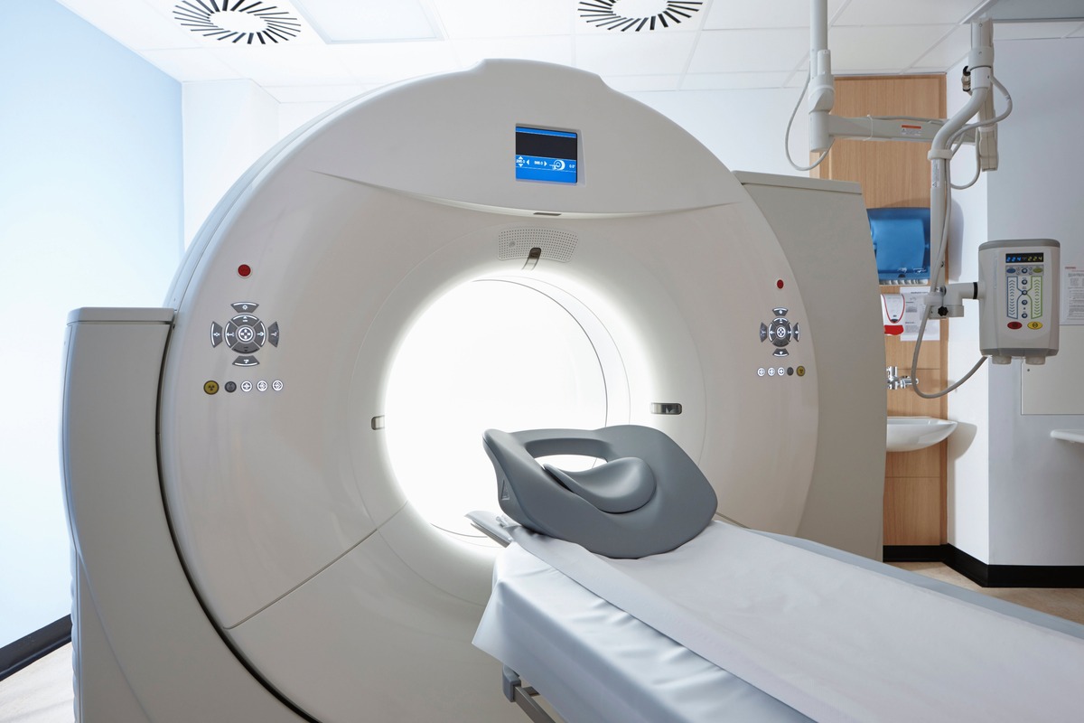
What is a CT scanner?
A CT (computed tomography) scanner is a powerful medical imaging device that combines X-ray technology with computer processing to create detailed cross-sectional images of various parts of the body. Unlike traditional X-rays that provide a 2D view, CT scanners generate a series of images taken from different angles, allowing physicians to examine structures layer by layer.
This revolutionary technology has transformed the field of radiology, enabling healthcare professionals to diagnose and treat a wide range of medical conditions with unprecedented accuracy. By providing detailed information about the body’s internal organs, bones, blood vessels, and tissues, CT scanners offer valuable insights that aid in early detection, monitoring, and treatment planning.
The functioning of a CT scanner involves X-ray beams passing through the body, and sophisticated detectors capturing the radiation that emerges on the other side. The scanner then uses advanced algorithms to analyze the data and reconstruct a detailed image, which can be viewed on a computer monitor or printed for further examination.
CT scans are beneficial in various medical scenarios, ranging from detecting tumors, fractures, and infections to guiding surgical interventions and monitoring treatment effectiveness. They are commonly used in fields like oncology, neurology, cardiology, and emergency medicine.
It’s worth noting that CT scans involve higher radiation levels compared to other imaging techniques, such as MRI or ultrasound. However, the benefits of the detailed information provided by CT scans often outweigh the associated risks, making it a valuable tool in clinical practice.
Over the years, CT scanners have evolved significantly, with advancements in technology leading to improved image quality, faster scan times, and reduced radiation exposure. This continuous innovation has made CT scanning an indispensable component of modern medical diagnostics.
The early days of medical imaging
Before the advent of CT scanners, medical imaging was limited to conventional X-ray machines, which provided only a limited view of the body’s internal structures. This posed significant challenges for accurate diagnosis and treatment planning.
The field of medical imaging started to emerge in the late 19th century when Wilhelm Conrad Roentgen discovered X-rays in 1895. This groundbreaking discovery paved the way for the development of imaging techniques that could capture internal images without invasive procedures.
Early X-ray machines used photographic plates to capture the X-ray images, which were time-consuming and involved a significant amount of radiation exposure for the patient. These limitations highlighted the need for a more efficient and safer imaging method.
As technology advanced, various imaging techniques were explored, such as fluoroscopy, which allowed real-time imaging of the body’s internal structures. However, these techniques still relied on X-ray technology and didn’t provide the level of detail needed for accurate diagnosis.
In the mid-20th century, the use of contrast agents, such as iodine-based dyes, started to enhance the visibility of certain anatomical structures on X-ray images. This breakthrough enabled better visualization of blood vessels and other soft tissues.
Despite these developments, there was still a significant gap in medical imaging technology. Radiologists and scientists recognized the need for a method that could provide detailed cross-sectional images of the body.
The concept of computed tomography (CT) imaging was born in the 1960s when a British engineer named Godfrey Hounsfield proposed using mathematical algorithms to reconstruct images from multiple X-ray projections. This marked a major turning point in medical imaging.
Researchers started experimenting with different approaches to implementing Hounsfield’s concept, but it wasn’t until 1971 that the first prototype of a CT scanner was built. The device utilized a moving X-ray source and detector to capture multiple images, which were then processed by a computer to create cross-sectional images.
This early CT scanner provided a significant advancement over traditional X-ray machines, allowing physicians to visualize internal structures in unprecedented detail. It was soon evident that CT scanning had the potential to revolutionize medical diagnostics.
The development of the CT scanner set the stage for a new era in medical imaging, laying the foundation for the remarkable advancements that we see in modern CT technology today.
The development of the CT scanner concept
The concept of the CT scanner, also known as computed tomography, began to take shape in the 1960s. It was a result of the pioneering work and collaboration of several scientists and engineers who aimed to overcome the limitations of traditional X-ray imaging.
One of the key figures in the development of the CT scanner concept was Godfrey Hounsfield, a British engineer. In 1967, he proposed using mathematical algorithms and computer processing to create cross-sectional images of the body. His idea was to use X-rays at different angles to capture multiple images that could then be reconstructed by a computer into detailed 2D and 3D images.
At the same time, Allan M. Cormack, a South African physicist, independently developed a mathematical method called the “Radon transform” to reconstruct images from X-ray projections. This method formed the basis for the reconstruction algorithms used in CT scanning.
Hounsfield and Cormack’s work laid the foundation for the development of the first CT scanner. However, the implementation of their ideas required advancements in computer technology and X-ray imaging techniques.
In 1971, Hounsfield and his team at EMI (Electrical and Musical Industries) in the UK built the first prototype of a CT scanner. This machine consisted of an X-ray tube that emitted a narrow X-ray beam, which was collimated into a series of fan-shaped beams. The detector on the opposite side measured the X-ray intensity after it passed through the body.
With the help of a computer, these measurements were fed into intricate algorithms to reconstruct cross-sectional images. The process was time-consuming, taking several hours to produce a single image, but it represented a groundbreaking advancement in medical imaging.
While Hounsfield was developing his prototype, another team led by Robert S. Ledley at Georgetown University Medical Center in the US was also working on a similar CT scanner concept. In 1973, they introduced their own prototype, which utilized a rotating X-ray source and detector to capture images at different angles.
These early CT scanners were large and complex machines, occupying entire rooms and requiring extensive technical expertise to operate. However, their potential to revolutionize medical diagnostics was evident from the beginning.
The development of the CT scanner concept was a significant milestone in medical imaging. It combined the power of X-ray technology with advanced computer algorithms, paving the way for a new era of diagnostic imaging that would transform medicine in ways previously unimaginable.
The first prototype of the CT scanner
After years of research and development, the concept of the CT scanner became a reality with the creation of the first working prototype. This pivotal moment in medical imaging history occurred in the early 1970s, marking a major breakthrough in diagnostic capabilities.
Godfrey Hounsfield, working at EMI in the United Kingdom, was instrumental in developing the first prototype of a CT scanner. In 1971, he successfully built a machine that could capture detailed cross-sectional images of the human body.
The initial CT scanner prototype was a large and complex device, consisting of an X-ray tube, a detector, and a computer system. The X-ray tube emitted a narrow beam of X-rays, which passed through the body and were detected by sensors on the other side. These detectors measured the intensity of the X-rays after they had interacted with the body’s tissues.
The captured data was then fed into a computer, and utilizing sophisticated algorithms, the computer processed the information to reconstruct detailed cross-sectional images. These images provided a unique view into the internal structures of the body, opening up new possibilities for medical diagnosis and treatment.
It’s important to note that the first prototype of the CT scanner had its limitations. It took several hours to produce a single image, as the data had to be processed meticulously by the computer. Despite these constraints, the potential of the technology was revolutionary, and researchers and medical professionals were eager to see its impact on patient care.
At around the same time, another team, led by Robert S. Ledley at Georgetown University Medical Center in the United States, also developed their own prototype of a CT scanner. Their device utilized a different approach, with a rotating X-ray source and detector to capture images from multiple angles. This provided more efficient data acquisition and quicker image reconstruction than Hounsfield’s prototype.
Although the first CT scanner prototypes exhibited limitations in terms of speed and image quality, they were significant milestones in medical imaging. They demonstrated the feasibility of the concept and paved the way for further development and refinement of the technology.
The successful creation of these early prototypes laid the foundation for the remarkable advancements that would occur in the field of CT scanning. Subsequent iterations of the CT scanner would address the limitations of the initial models, resulting in faster scan times, improved image quality, and enhanced patient comfort.
The first prototype of the CT scanner marked a turning point in medical imaging, opening up a new era of diagnostic capabilities and transforming the way medical professionals could visualize and understand the human body.
The momentous breakthrough of the CT scanner
The development of the CT scanner brought about a momentous breakthrough in the field of medical imaging. This groundbreaking technology revolutionized diagnostic capabilities and had a profound impact on patient care and treatment.
The introduction of the CT scanner provided a significant leap forward in medical imaging, as it allowed physicians to obtain detailed and precise cross-sectional images of the body’s internal structures. This level of visualization was far superior to what was previously possible with traditional X-ray techniques.
With the ability to capture images from multiple angles, the CT scanner provided a three-dimensional view of organs, bones, blood vessels, and other tissues. This enabled medical professionals to detect abnormalities, diagnose diseases, plan surgeries, and monitor treatment progress with unprecedented accuracy.
Prior to the CT scanner, diagnosis and treatment decisions were often based on limited information obtained from X-rays, physical examinations, and patient symptoms. The CT scanner transformed medical practice by providing detailed anatomical information, allowing for more targeted and effective interventions.
One of the key advantages of the CT scanner was its ability to distinguish between different types of soft tissues, providing greater insight into the body’s internal structures. This made it particularly valuable in identifying tumors, evaluating organ functionality, and diagnosing conditions such as strokes and cardiovascular diseases.
Furthermore, the CT scanner played a crucial role in the early detection of various types of cancer. By providing a clear view of abnormal growths or masses, it allowed for timely interventions and improved patient outcomes.
The impact of the CT scanner on medical practice was profound, as it helped reduce the need for invasive procedures such as exploratory surgeries. Instead, physicians could rely on the detailed information obtained from CT scans to guide their diagnostic and treatment decisions.
Not only did the CT scanner revolutionize diagnostic capabilities, but it also helped pave the way for minimally invasive procedures. By accurately mapping out the anatomy of the body, surgeons could plan and navigate complex procedures with precision, resulting in reduced risks and faster recovery times for patients.
Overall, the introduction of the CT scanner was a momentous breakthrough in the field of medical imaging. Its ability to provide detailed cross-sectional images transformed medical practice, enabling early detection, accurate diagnosis, and effective treatment of a wide range of conditions.
Public release and commercialization of the CT scanner
After the successful development of the first CT scanner prototypes, the next step was to bring this revolutionary technology to the medical community and make it commercially available. The public release and commercialization of the CT scanner marked a significant milestone in the history of medical imaging.
In 1972, the first clinical installation of a CT scanner took place at Atkinson Morley’s Hospital in London, UK. This marked the beginning of a new era in diagnostic imaging as medical professionals gained access to this groundbreaking technology.
The release of the CT scanner ignited immense interest and excitement within the medical community. Radiologists, surgeons, and physicians recognized the potential of this advanced imaging technology and its ability to enhance patient care.
Commercialization of the CT scanner came soon after its initial introduction. Companies such as EMI (Electrical and Musical Industries) and General Electric (GE) played significant roles in refining and manufacturing CT scanners for widespread use.
Over time, advancements in computer technology and imaging techniques led to the development of faster and more sophisticated CT scanners. These new generations of scanners offered improved image quality, reduced scan times, and enhanced patient comfort, making them even more attractive to healthcare institutions.
The commercial availability of CT scanners also coincided with the increasing demand for medical imaging services. As more medical facilities recognized the benefits of CT scanning, they began investing in these devices to enhance their diagnostic capabilities.
As the popularity of CT scanners grew, so did their accessibility. More hospitals and clinics were equipped with CT scanners, enabling a wider range of patients to benefit from this advanced imaging technology. The commercialization of the CT scanner made it an integral part of medical practice in the diagnosis and treatment of various conditions.
Alongside the increased availability of CT scanners, protocols and guidelines were developed to ensure the safe and effective use of this technology. Radiation safety measures and specific imaging techniques were established to optimize patient care and minimize radiation exposure.
The commercialization of the CT scanner not only revolutionized medical diagnostics but also had a significant impact on medical education and research. Radiologists and other healthcare professionals were now able to explore the intricacies of the human body in far greater detail, leading to new discoveries and advancements in medical knowledge.
The public release and commercialization of the CT scanner marked a turning point in medical imaging. This advanced technology became widely accessible, transforming the field of diagnostic imaging and empowering healthcare professionals to provide better and more precise care to their patients.
The impact of the CT scanner on medicine
The introduction of the CT scanner has had a profound impact on the field of medicine, revolutionizing diagnostic capabilities and transforming patient care. This advanced imaging technology has opened up new possibilities for early detection, accurate diagnosis, treatment planning, and monitoring of various medical conditions.
One of the key impacts of the CT scanner is its ability to provide detailed cross-sectional images of the body’s internal structures. This has significantly improved the accuracy and precision of diagnoses across a wide range of medical specialties, including radiology, oncology, neurology, and cardiology.
By offering three-dimensional views of organs, tissues, and blood vessels, CT scans allow physicians to identify abnormalities, such as tumors, fractures, and infections, with remarkable precision. This early detection enables prompt intervention and treatment, leading to improved patient outcomes.
Moreover, the CT scanner plays a crucial role in treatment planning for various conditions. Surgeons can use CT scans to accurately map out the anatomy of the body, aiding in surgical navigation and ensuring precise interventions. CT scans also guide radiation therapy, allowing for the targeted delivery of radiation to cancerous tissues while minimizing damage to nearby healthy tissues.
The impact of the CT scanner on emergency medicine cannot be overstated. In critical situations, such as trauma cases or acute strokes, CT scans provide rapid and crucial information that enables physicians to make critical decisions regarding interventions and treatment options. This timely information saves lives and enhances patient outcomes.
Another significant impact of the CT scanner is the reduction in the need for invasive procedures. Prior to its introduction, invasive exploratory surgeries were often required to diagnose certain conditions. With the detailed imaging provided by CT scans, physicians can now make accurate diagnoses without the need for invasive interventions, reducing patient discomfort, risks, and recovery times.
Additionally, the CT scanner has had a profound impact on medical research and education. The detailed images obtained from CT scans have contributed to medical knowledge, allowing researchers to study the human body in ways that were previously not possible. Medical students and trainees also benefit from the ability to observe and learn from these detailed images, enhancing their understanding of anatomy and pathology.
With continuous advancements in CT scanning technology, such as faster scan times, improved image quality, and reduced radiation exposure, the impact of CT scanners on medicine is expected to continue expanding. It has become an indispensable tool in the diagnosis, treatment, and monitoring of a broad range of medical conditions, improving patient care and advancing medical knowledge.
Technological advancements in CT scanning over the years
Since its introduction, CT scanning technology has undergone significant advancements, resulting in improved image quality, faster scan times, and enhanced patient comfort. These technological innovations have revolutionized the field of diagnostic imaging, allowing for more accurate and efficient diagnoses of various medical conditions.
One of the major advancements in CT scanning technology is the development of multi-slice or multi-detector CT scanners. These scanners feature multiple detector arrays that can acquire data from multiple slices of the body simultaneously. This not only significantly reduces scan times but also improves image resolution and enables the visualization of smaller structures.
The use of faster rotation speeds in modern CT scanners has also contributed to shorter scan times. With faster gantry rotations, more images can be obtained within a shorter period, facilitating efficient patient throughput while maintaining image quality.
Another significant advancement is the introduction of spiral or helical CT scanning. This technique involves continuous gantry rotation while the patient is moved through the scanner, resulting in a helical or spiral trajectory of the X-ray beam. This allows for faster acquisition of volumetric data, reducing motion artifacts and improving image quality, particularly in areas prone to motion, such as the heart and lungs.
Computed Tomography Angiography (CTA) is another important development in CT scanning technology. CTA combines the use of intravenous contrast agents with CT scanning to visualize blood vessels and evaluate vascular anatomy. This has revolutionized the detection and diagnosis of vascular diseases such as arterial blockages, aneurysms, and pulmonary embolisms.
Advancements in reconstruction algorithms have also played a significant role in improving the quality of CT images. Iterative reconstruction algorithms, for example, use mathematical models to iteratively refine the reconstructed images, reducing image noise and enhancing image sharpness.
The advent of low-dose CT scanning has been a major breakthrough in patient radiation dose reduction. Utilizing advanced techniques such as tube current modulation and automatic exposure control, low-dose CT imaging optimizes the radiation dose delivered to the patient while maintaining image quality, contributing to patient safety.
Recent advancements in CT scanning technology have also focused on reducing the use of iodinated contrast agents, particularly for patients with kidney dysfunction or allergies. Techniques such as Dual Energy CT and Spectral CT provide information about tissue composition and perfusion, reducing the reliance on contrast agents in certain situations.
The integration of artificial intelligence (AI) and machine learning algorithms into CT scanners is another significant development. AI algorithms have the potential to automate image analysis, assist in precise diagnosis, and improve workflow efficiency. They can aid in the detection and characterization of abnormalities, streamline image interpretation, and enhance clinical decision-making.
These technological advancements in CT scanning have greatly improved patient care, allowing for more accurate diagnoses, faster scanning, reduced patient radiation exposure, and enhanced overall imaging capabilities. As technology continues to evolve, CT scanners will continue to play a pivotal role in the diagnosis and treatment of various medical conditions, further enhancing the field of diagnostic imaging.
Current state of CT scanning technology
The field of CT scanning technology continues to evolve and innovate, leading to advancements that enhance image quality, increase scan speed, reduce radiation dose, and improve overall patient experience. The current state of CT scanning technology reflects the ongoing commitment to improving diagnostic capabilities and patient care.
One significant development in the current state of CT scanning is the widespread adoption of advanced imaging techniques, such as Dual Energy CT and Spectral CT. These techniques provide additional information about tissue composition, perfusion, and material identification, leading to more accurate diagnoses and treatment planning.
Another notable advancement is the growing use of artificial intelligence (AI) and machine learning algorithms in CT scanning. AI algorithms can assist in image interpretation, automate certain tasks, and provide quantitative analysis to aid in diagnosing and characterizing abnormalities. This integration of AI into CT scanners has the potential to enhance efficiency and improve diagnostic accuracy.
Low-dose CT imaging has become a standard practice in many healthcare institutions, with advanced dose reduction techniques implemented to minimize radiation exposure without compromising image quality. This focus on patient safety has resulted in significant advancements in dose modulation, iterative reconstruction algorithms, and other dose-reduction strategies.
Advancements in detector technology have also contributed to improvements in image quality and diagnostic capabilities. The use of more sensitive and efficient detectors allows for better spatial resolution, reduced image noise, and enhanced visualization of smaller structures.
The integration of CT scanners with other imaging modalities, such as positron emission tomography (PET) or magnetic resonance imaging (MRI), has also gained momentum in recent years. These hybrid imaging systems provide comprehensive and synergistic information, enabling more accurate diagnoses, treatment planning, and monitoring of complex conditions.
Additionally, advancements in patient-centric design have improved the overall experience for individuals undergoing CT scans. Innovations in gantry design, patient positioning, and noise reduction techniques aim to enhance patient comfort and reduce anxiety during the imaging process.
Modern CT scanners are equipped with user-friendly interfaces, intuitive software, and advanced automation features to streamline workflow and improve efficiency. These advancements allow for faster image acquisition, reconstruction, and analysis, facilitating more timely diagnoses and treatment decisions.
Furthermore, the current state of CT scanning technology emphasizes interoperability and integration with picture archiving and communication systems (PACS) and electronic health records (EHR). This seamless integration of imaging data and patient information enhances communication and collaboration among healthcare professionals, ultimately improving patient care.
Looking ahead, the future of CT scanning technology holds promise for continued advancements in areas such as image quality, speed, accessibility, and clinical applications. As technology evolves, CT scanners will remain an instrumental tool in medical diagnostics, enabling early detection, precise interventions, and improved patient outcomes.