
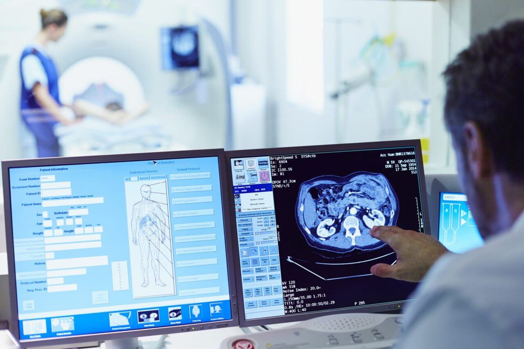
CT Scan
A CT scan, which stands for computed tomography, is a diagnostic imaging technique commonly used to view a patient’s internal organs. It utilizes a combination of X-rays and computer technology to create cross-sectional images of the body. The CT scanner rotates around the patient and captures multiple X-ray images from different angles. These images are then processed by a computer to generate detailed 3D images.
CT scans offer a high level of precision and clarity, making them an invaluable tool in diagnosing various medical conditions. They can be used to examine organs such as the brain, chest, abdomen, pelvis, and bones. CT scans are particularly useful in detecting tumors, studying blood vessels, evaluating injuries, and guiding medical procedures.
During a CT scan, the patient lies on a table that moves through the scanner. It is essential to remain still during the procedure to avoid blurring the images. In some cases, a contrast dye may be administered intravenously to enhance the visibility of certain structures or conditions.
One of the significant advantages of CT scans is their speed. They can provide detailed images quickly, reducing the time patients need to spend in the scanner. Additionally, CT scans are generally non-invasive and painless. However, as they involve exposure to ionizing radiation, precautions are taken to minimize radiation exposure, especially in pregnant women and children.
CT scans have revolutionized medical imaging, allowing physicians to make accurate diagnoses and develop appropriate treatment plans. They play a crucial role in detecting abnormalities, planning surgeries, monitoring disease progression, and assessing the effectiveness of treatment. The detailed and comprehensive information provided by CT scans helps medical professionals make informed decisions, leading to improved patient outcomes.
MRI Scan
An MRI scan, short for magnetic resonance imaging, is a widely used medical imaging technique that provides detailed images of a patient’s internal organs and tissues. Unlike X-rays or CT scans, MRI scans use a strong magnetic field and radio waves to generate these images, making them particularly useful for capturing soft tissue structures.
During an MRI scan, the patient lies on a table that slides into a large cylindrical machine. The machine creates a magnetic field that causes the body’s hydrogen atoms to align in a specific way. Radio waves are then used to create signals that are processed by a computer to generate highly detailed images of the body’s structures.
MRI scans can be performed on various parts of the body, including the brain, spine, joints, abdomen, and pelvis. They are particularly valuable for diagnosing conditions such as tumors, spinal cord injuries, joint abnormalities, and neurological disorders. MRI scans can also provide valuable information about blood flow and help identify vascular conditions.
One significant advantage of MRI scans is that they do not expose patients to ionizing radiation. This makes them a safe option, especially for those who require multiple imaging procedures or for young children. However, there are some limitations to consider. Patients with certain metal implants, such as pacemakers or cochlear implants, may not be eligible for an MRI scan due to safety concerns.
Another advantage of MRI scans is their ability to provide detailed multi-planar images, allowing doctors to view structures from different angles. This three-dimensional information is valuable for accurate diagnosis and treatment planning. In some cases, contrast agents may be used to enhance the visibility of specific tissue or conditions during the MRI scan.
Overall, MRI scans are a versatile and powerful tool in modern medicine. They provide essential information for diagnosing a wide range of medical conditions and guiding treatment decisions. The detailed images produced by MRI scans can help physicians visualize even the subtlest abnormalities, leading to more accurate diagnoses and improved patient care.
Ultrasound
Ultrasound imaging, commonly known as sonography, is a non-invasive diagnostic technique that uses high-frequency sound waves to produce real-time images of a patient’s internal organs. It is a safe and painless procedure that is widely used in various medical specialties, including obstetrics, cardiology, and radiology.
During an ultrasound examination, a handheld device called a transducer is placed on the patient’s skin. The transducer emits sound waves that bounce off the internal organs and tissues, creating echoes. These echoes are then converted into visual images by a computer, providing valuable information about the structure and function of the organs being examined.
Ultrasound scans are commonly associated with pregnancy as they are frequently used to monitor the development of the fetus. They can provide detailed images of the baby’s growth, assess the health of the placenta, and detect any abnormalities. In addition to obstetrics, ultrasound is used for evaluating the liver, kidneys, heart, gallbladder, and other organs.
One of the major advantages of ultrasound is its safety. It does not involve the use of ionizing radiation, making it suitable for all age groups, including infants and pregnant women. Additionally, ultrasound scans can be performed in real-time, allowing doctors to observe moving structures, monitor blood flow, and assess organ functions dynamically.
Another significant benefit of ultrasound is its portability and accessibility. Compared to other imaging techniques, ultrasound machines are smaller, more affordable, and can be easily transported. This makes ultrasound a valuable tool in emergency situations, where immediate assessment of internal organs may be vital.
Ultrasound scans are typically performed by skilled sonographers who can interpret the images and provide valuable diagnostic information to the healthcare team. In some cases, a specialized type of ultrasound called Doppler ultrasound may be used to assess blood flow or detect inflammation or blockages in blood vessels.
Overall, ultrasound imaging is a versatile and safe diagnostic tool that provides valuable insights into a patient’s internal organs and structures. Its real-time capabilities, portability, and lack of radiation exposure make it an essential component of modern medical practice.
X-Ray
X-ray imaging, also known as radiography, is one of the oldest and most commonly used medical imaging techniques. It involves using small amounts of ionizing radiation to create images of a patient’s internal structures, including bones, lungs, and certain soft tissues.
During an X-ray, a focused beam of X-ray radiation is passed through the body. The X-ray machine detects the radiation that passes through the body and captures the resulting image on a digital detector or a traditional X-ray film. Dense structures like bones appear white on the X-ray image, while less dense tissues and organs appear in shades of gray.
X-rays are particularly useful for diagnosing fractures, tumors, infections, and abnormalities in the chest or abdomen. The images produced by X-rays can help physicians identify injuries, visualize changes in bone density, and spot certain diseases at an early stage.
One of the primary advantages of X-ray imaging is its speed and simplicity. X-ray exams are generally quick, taking only a few minutes, which is especially beneficial in emergency situations where prompt diagnosis is crucial. Furthermore, X-ray equipment is widely available in hospitals and clinics, making it a cost-effective and accessible imaging option.
However, it’s important to note that X-rays involve exposure to ionizing radiation, which can have potential risks. Healthcare professionals take precautions to limit radiation exposure by using lead aprons and collimation techniques to focus the X-ray beam only on the area of interest. Special care is also taken with pregnant women, where the benefits of an X-ray must outweigh the potential risks to the fetus.
Advancements in technology have led to digital X-ray imaging, which offers several advantages over traditional film-based X-rays. Digital X-rays provide instant results, allow for image manipulation for enhanced visualization, and reduce the need for chemicals used in film development.
In recent years, X-ray imaging has also become more advanced with the introduction of specialized techniques, such as fluoroscopy and computed tomography (CT). Fluoroscopy uses continuous X-rays to visualize moving structures, while CT scans provide detailed 3D images of the body.
Endoscopy
Endoscopy is a medical procedure that allows doctors to visually examine and diagnose conditions within the body’s internal organs and cavities using a flexible, lighted instrument called an endoscope. This minimally invasive technique has revolutionized diagnostic and therapeutic procedures in various medical fields, including gastroenterology, pulmonology, and urology.
During an endoscopy, the endoscope is inserted through a natural opening in the body or a small incision. The endoscope contains a light source and a camera that captures high-definition images of the internal organs and structures. These images are displayed on a monitor, allowing the medical team to assess the condition of the tissues and perform necessary procedures.
Endoscopy enables physicians to directly visualize areas of concern, such as the gastrointestinal tract, respiratory system, urinary tract, and reproductive organs. It can help diagnose conditions such as ulcers, polyps, tumors, infections, and abnormalities in these areas.
One of the most common types of endoscopy is gastroscopy, which involves examining the esophagus, stomach, and upper part of the small intestine. It is useful for diagnosing conditions like gastroesophageal reflux disease (GERD), gastritis, and peptic ulcers.
Another commonly performed endoscopic procedure is colonoscopy, which allows the examination of the large intestine or colon. It helps in the detection and removal of polyps, screening for colorectal cancer, and diagnosis of inflammatory bowel disease (IBD).
Endoscopy offers several advantages over traditional surgical procedures. Since it is minimally invasive, it reduces the risks and complications associated with open surgeries. Patients typically experience less pain, have shorter recovery times, and require shorter hospital stays compared to surgical interventions.
Moreover, endoscopy often eliminates the need for more invasive diagnostic tests, such as exploratory surgery, by providing direct visualization and biopsies of affected areas. In some cases, therapeutic procedures can be performed during endoscopy, such as the removal of polyps, dilation of strictures, or placement of stents.
Although endoscopy is generally safe, there is a slight risk of complications such as bleeding, infection, or perforation. However, these risks are rare and closely monitored by the medical team.
Overall, endoscopy has significantly improved diagnostic capabilities and treatment outcomes in various medical specialties. By providing a direct visualization of internal organs, it allows for timely and accurate diagnoses, leading to appropriate treatment plans and improved patient care.
PET Scan
Positron emission tomography, commonly known as a PET scan, is a specialized imaging technique that provides detailed information about the metabolic activity of cells in the body. It is a valuable diagnostic tool in oncology, neurology, and cardiology, as it allows for the detection and evaluation of various diseases and conditions.
PET scans involve the injection of a small amount of radioactive material, known as a radiotracer, into the patient’s bloodstream. The radiotracer emits positrons, which are detected by the PET scanner. These positrons collide with electrons in the body, resulting in the production of gamma ray photons. The PET scanner measures the distribution of these photons, creating images that reflect the metabolic activity of cells.
PET scans are particularly useful in the detection and staging of cancers. Cancer cells have a higher metabolic rate than normal cells, which can be detected by the increased uptake of the radiotracer. PET scans can help identify the location, size, and extent of tumors, as well as evaluate the response to treatment.
In addition to oncology, PET scans can also provide valuable information in neurology. They can help diagnose and differentiate various neurodegenerative diseases, such as Alzheimer’s disease and Parkinson’s disease. PET scans can also assess brain function and identify areas of abnormal activity in conditions such as epilepsy and brain tumors.
PET scans can evaluate the blood flow and viability of heart tissue, making them useful in cardiology. They can assess cardiac function, myocardial perfusion, and identify regions of damaged or scarred tissue. PET scans can assist in the diagnosis and risk stratification of coronary artery disease and guide the planning of interventions such as angioplasty or bypass surgery.
One of the advantages of PET scans is their ability to provide functional information. They can demonstrate how organs and tissues are functioning at a cellular level, allowing for more accurate diagnoses and treatment planning. PET scans can also be combined with other imaging modalities, such as CT or MRI, to provide a more comprehensive evaluation.
It’s important to note that PET scans involve exposure to ionizing radiation due to the use of the radiotracer. However, the amount of radiation used in PET scans is considered safe and does not pose significant health risks.
Overall, PET scans play a crucial role in the diagnosis, staging, and monitoring of various diseases. By providing information about the metabolic activity of cells, they offer valuable insights into the underlying processes and help guide appropriate treatment decisions.