
Diagnostic Imaging
Diagnostic imaging is one of the primary uses of ultrasound technology. It involves the use of high-frequency sound waves to create images of the internal structures of the body. This non-invasive and painless procedure helps doctors visualize and diagnose various medical conditions.
Ultrasound imaging is commonly used in different medical specialties for diagnostic purposes. It allows healthcare professionals to examine organs, tissues, and blood flow in real-time, providing valuable insights into the patient’s health. Here are some of the key applications of ultrasound in diagnostic imaging:
- Abdominal Imaging: Ultrasound scans are frequently used to assess the organs in the abdomen, including the liver, gallbladder, pancreas, kidneys, and spleen. It is useful in detecting abnormalities such as tumors, cysts, stones, and inflammation.
- Obstetrics and Gynecology: Ultrasound is widely employed to monitor the development and well-being of the fetus during pregnancy. It helps determine the gestational age, detect any problems with fetal growth or positioning, and diagnose conditions like ectopic pregnancy or multiple pregnancies.
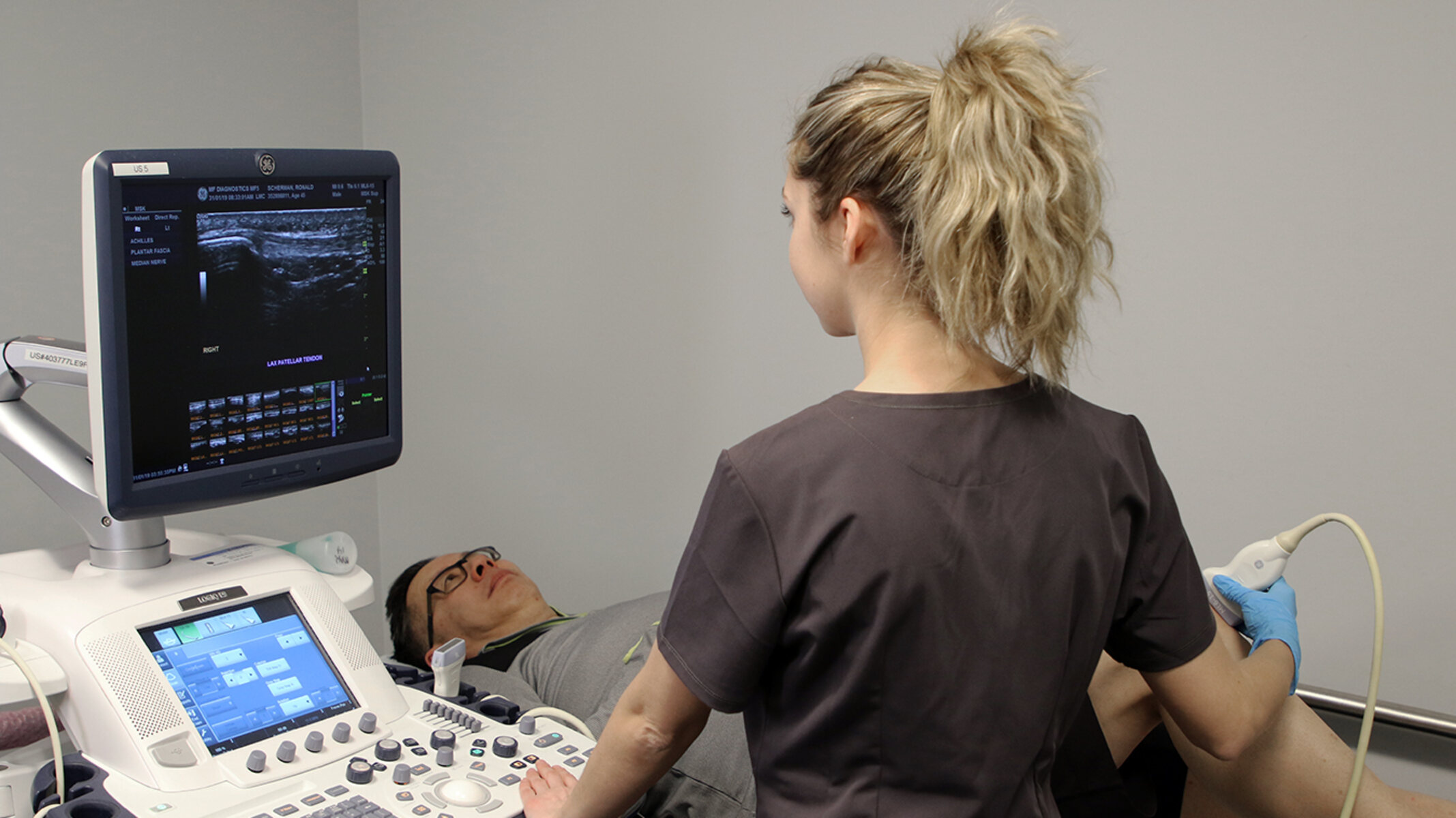
- Musculoskeletal Imaging: Ultrasound plays a vital role in evaluating soft tissues, muscles, tendons, ligaments, and joints. It assists in diagnosing various musculoskeletal conditions, such as sprains, strains, tears, and arthritis.
- Cardiovascular Imaging: Ultrasound is extensively used for assessing heart health. It helps in visualizing the structure and function of the heart, including the chambers, valves, and blood vessels. Echocardiography, a type of ultrasound, aids in diagnosing and monitoring heart diseases.
- Breast Imaging: Ultrasound is commonly used alongside mammography to examine breast tissue. It helps differentiate between benign and malignant masses, guide breast biopsies, and monitor breast conditions.
Ultrasound imaging offers numerous advantages, including its non-invasiveness, portability, and absence of ionizing radiation. However, it does have limitations, such as its dependency on operator skill, body habitus, and the presence of air or gas obscuring the imaging. Overall, diagnostic imaging with ultrasound continues to revolutionize healthcare by providing valuable diagnostic information and improving patient outcomes.
Obstetrics and Gynecology
Obstetrics and gynecology is a medical specialty that focuses on the health of the female reproductive system, including pregnancy, childbirth, and the diagnosis and treatment of reproductive disorders. Ultrasound technology plays a crucial role in obstetrics and gynecology, offering valuable insights into the health and well-being of both the mother and the fetus.
In obstetrics, ultrasound is used extensively to monitor the progress and development of the fetus during pregnancy. It allows healthcare professionals to determine the gestational age, assess fetal growth, and detect any potential abnormalities or complications. Ultrasound imaging can provide detailed images of the fetus, including its anatomy, organs, and movements. This information helps doctors ensure the well-being of the baby and make informed decisions regarding prenatal care.
Gynecology also benefits from the use of ultrasound technology. It aids in the diagnosis and evaluation of various gynecological conditions. For instance, ultrasound can help identify uterine fibroids, ovarian cysts, and other structural or functional abnormalities. It is used for evaluating the endometrium, assessing the thickness of the uterine lining, and detecting any abnormalities or signs of disease. Ultrasound is also employed during fertility treatments, assisting in monitoring the response to fertility drugs, tracking egg development, and guiding procedures like egg retrieval or embryo transfer.
Additionally, ultrasound can be used for procedures such as hysterosonography, which involves injecting saline into the uterus to get a clearer image of the uterine cavity; and ultrasound-guided biopsies, where a small tissue sample is obtained for further analysis.
The use of ultrasound in obstetrics and gynecology has revolutionized prenatal care and women’s healthcare. Its non-invasive nature, real-time imaging capabilities, and absence of radiation make it a preferred diagnostic tool. Ultrasound allows for early detection of fetal abnormalities, helps manage high-risk pregnancies, and guides medical interventions when necessary. In gynecology, it offers a safe and accurate method of evaluating the reproductive system, aiding in the diagnosis and treatment of various conditions.
Overall, ultrasound technology continues to be a cornerstone in obstetrics and gynecology, empowering healthcare professionals to provide quality care, ensure healthy pregnancies, and improve the overall reproductive health of women.
Cardiology
Cardiology is the branch of medicine that deals with the study, diagnosis, and treatment of heart diseases and disorders. Ultrasound technology, specifically echocardiography, plays a vital role in cardiology, allowing doctors to examine the structure and function of the heart non-invasively.
Echocardiography uses ultrasound waves to create detailed images of the heart. It provides valuable information about the heart’s chambers, valves, blood flow, and overall cardiac function. This imaging modality helps in the diagnosis and monitoring of various cardiovascular conditions.
One of the primary applications of echocardiography is in assessing heart function. It allows healthcare professionals to measure the ejection fraction, which is a measure of the heart’s pumping ability. Echocardiography can detect abnormalities in the heart’s size, shape, and wall thickness, providing insights into conditions like cardiomyopathy, heart failure, and congenital heart defects.
Echocardiography is also used in the evaluation of heart valve disorders. It can assess the movement and function of heart valves and detect conditions such as valve stenosis or regurgitation. This information helps guide treatment decisions, such as the need for valve repair or replacement.
Furthermore, echocardiography plays a crucial role in diagnosing and managing coronary artery disease. It can identify areas of reduced blood flow to the heart muscle, evaluate the extent of blockages in the coronary arteries, and assess the overall perfusion of the heart. This information is valuable for determining the need for interventions like angioplasty or coronary bypass surgery.
Another important use of echocardiography is in evaluating the presence of blood clots within the heart chambers. It helps in the detection of conditions like atrial fibrillation and provides information on the risk of stroke.
The versatility of echocardiography extends to pediatric cardiology as well, aiding in the diagnosis and management of congenital heart defects in children.
In summary, echocardiography, a form of ultrasound imaging, plays a critical role in the field of cardiology. It provides a safe, non-invasive, and detailed assessment of the heart’s structure and function, aiding in the diagnosis, treatment, and monitoring of various cardiovascular conditions. Its ability to provide real-time images and measurements makes it an invaluable tool in the field of cardiology.
Ophthalmology
Ophthalmology is the branch of medicine that specializes in the study, diagnosis, and treatment of eye diseases and disorders. Ultrasound technology plays a significant role in ophthalmology by providing valuable insights into the structures of the eye that are not easily visible through other imaging modalities.
Ultrasound imaging in ophthalmology is particularly useful when the view of the eye is obstructed or when traditional methods of imaging, such as optical coherence tomography (OCT), are not feasible. This non-invasive and painless procedure uses high-frequency sound waves to generate images of the eye’s internal structures, including the retina, vitreous, and optic nerve.
One of the primary applications of ultrasound in ophthalmology is in the evaluation of ocular trauma or eye emergencies. When visualizing the eye is difficult due to swelling, bleeding, or other obstructions, ultrasound can provide valuable information about the extent of damage, the presence of foreign bodies, or the integrity of ocular structures.
Ultrasound imaging is also used in the diagnosis and management of retinal detachments. It helps ophthalmologists visualize and assess the position and extent of the detached retina, aiding in surgical planning and decision-making.
In addition to trauma and retinal detachments, ultrasound is commonly used in ophthalmology for the evaluation of intraocular tumors. It helps determine the size, location, and characteristics of tumors within the eye, assisting ophthalmologists in diagnosis, treatment planning, and monitoring of tumor progression.
Another application of ultrasound in ophthalmology is the assessment of the optic nerve. This non-invasive technique can provide valuable information about the size, shape, and patency of the optic nerve, aiding in the diagnosis of conditions such as optic neuritis, optic nerve head drusen, or optic nerve tumors.
In summary, ultrasound technology plays a crucial role in ophthalmology by providing detailed imaging of ocular structures, especially when traditional methods are limited. It aids in the assessment of ocular trauma, evaluation of retinal detachments, diagnosis of intraocular tumors, and evaluation of the optic nerve. The non-invasive nature of ultrasound imaging makes it a valuable tool for ophthalmologists in the diagnosis and management of various eye conditions.
Dermatology
Dermatology is the medical specialty that focuses on the diagnosis and treatment of diseases related to the skin, hair, and nails. While dermatologists primarily rely on visual examination and other diagnostic techniques, ultrasound technology has emerged as a valuable tool in the field of dermatology, offering additional insights into skin conditions and aiding in treatment planning.
Ultrasound imaging in dermatology provides a non-invasive and real-time assessment of the skin’s deeper layers, which is not easily visible with the naked eye. It is particularly useful in evaluating skin lesions, guiding procedures, and monitoring treatment progress.
One of the key applications of ultrasound in dermatology is in the assessment of skin tumors and masses. It helps evaluate the depth, borders, and characteristics of the lesion, aiding in determining its nature and guiding decisions about appropriate treatment options. Ultrasound is beneficial in distinguishing between benign and malignant tumors, assisting dermatologists in biopsy planning and surgical management.
Additionally, ultrasound technology has proven valuable in the evaluation of inflammatory skin conditions, such as abscesses or cellulitis. It helps determine the extent of involvement, ruling out deeper involvement or the presence of complications like collections or fistulas. This information guides appropriate treatment strategies and helps dermatologists provide optimal care to their patients.
Moreover, ultrasound is increasingly being used to guide minimally invasive procedures in dermatology. It assists in localizing the target area, improving the accuracy of injections, and reducing the risk of complications. Ultrasound-guided procedures like biopsies, cyst aspirations, and introduction of therapeutic agents into specific layers of the skin have become more precise and effective with the aid of ultrasound imaging.
The use of ultrasound in dermatology offers several advantages, including its ability to provide real-time imaging, its non-invasive nature, and its portability. It allows dermatologists to visualize structures beneath the skin surface, providing valuable diagnostic information and aiding in treatment decisions.
In summary, the incorporation of ultrasound technology in dermatology has enhanced the diagnostic capabilities and treatment options available to dermatologists. It aids in the evaluation of skin lesions, assists in the diagnosis of skin tumors, guides procedures, and improves the overall management of various dermatological conditions. The non-invasive and real-time nature of ultrasound imaging has revolutionized the field of dermatology, empowering dermatologists to provide accurate diagnoses and effective treatments for their patients.
Orthopedics
Orthopedics is a medical specialty that focuses on the diagnosis, treatment, and prevention of conditions and injuries related to the musculoskeletal system, including bones, joints, tendons, ligaments, and muscles. Ultrasound technology plays a significant role in orthopedics, providing valuable imaging capabilities that aid in the assessment and management of various musculoskeletal conditions.
One of the primary applications of ultrasound in orthopedics is in the evaluation of soft tissue structures, such as tendons and ligaments. It allows healthcare professionals to assess the integrity, thickness, and overall condition of these structures. Ultrasound imaging is particularly valuable in diagnosing conditions such as tendonitis, ligament tears, and muscle strains. It aids in determining the extent of injury, guiding treatment decisions, and monitoring the healing process.
Furthermore, ultrasound is commonly used in orthopedics to guide various procedures, including injections and aspirations. Ultrasound-guided injections provide precise targeting of affected areas, increasing the accuracy and effectiveness of the treatment. It allows for visual confirmation of the needle placement, minimizing the risk of complications and enhancing patient outcomes.
Ultrasound imaging is also employed in the assessment of joint conditions, such as arthritis or synovitis. It helps in visualizing joint spaces, assessing the presence of inflammation, and evaluating joint fluid. This information aids in the diagnosis and treatment planning for various joint disorders.
In addition to evaluating soft tissues and joints, ultrasound technology has proven useful in assessing fractures and evaluating healing progress. It aids in visualizing the fracture line, confirming alignment, and monitoring the healing process. Ultrasound-guided procedures, such as fracture reductions or aspirations, can be performed with real-time imaging guidance, improving accuracy and outcomes.
The use of ultrasound in orthopedics offers several advantages, including its non-invasiveness, real-time imaging capabilities, and absence of ionizing radiation. It allows for dynamic assessment, as the musculoskeletal structures can be evaluated during movement. Ultrasound is also portable and readily accessible, making it a versatile tool in orthopedic clinics and settings.
In summary, ultrasound technology has become an invaluable tool in orthopedics, aiding in the evaluation, diagnosis, and management of various musculoskeletal conditions. It provides valuable imaging of soft tissues, assists in guiding procedures, and aids in the assessment of fractures and joint disorders. The non-invasive nature and real-time capabilities of ultrasound make it an essential component of modern orthopedic practice, enhancing patient care and outcomes.
Urology
Urology is a medical specialty that focuses on the diagnosis and treatment of conditions related to the urinary tract and the male reproductive system. Ultrasound technology plays a crucial role in urology, providing valuable imaging capabilities that aid in the assessment, diagnosis, and management of various urologic conditions.
One of the primary applications of ultrasound in urology is the evaluation of the kidneys. It allows healthcare professionals to visualize the size, shape, and position of the kidneys, as well as detect any abnormalities such as kidney stones, cysts, or tumors. Ultrasound imaging is particularly useful in assessing kidney function by measuring the size of the renal arteries and veins and evaluating blood flow to the kidneys.
Moreover, ultrasound is commonly used to assess the urinary bladder. It helps in determining the capacity and presence of any structural abnormalities, such as bladder stones or tumors. Ultrasound imaging is also valuable in guiding procedures such as bladder catheterization or bladder biopsies.
In addition to the kidneys and bladder, ultrasound plays a vital role in various other urologic applications. It aids in the evaluation of the prostate gland, assisting in the diagnosis of prostate enlargement (benign prostatic hyperplasia) or prostate cancer. Ultrasound-guided prostate biopsies are commonly performed to obtain tissue samples for further analysis.
Furthermore, ultrasound imaging is used to assess the male reproductive system. It aids in the assessment of the testicles, aiding in the diagnosis of conditions such as testicular torsion, varicocele, or tumors. Ultrasound can also be used to evaluate the scrotum and surrounding structures, providing valuable information for surgical planning or interventions.
The use of ultrasound in urology offers numerous advantages. It is a non-invasive, painless, and radiation-free imaging technique. It provides real-time imaging, allowing for dynamic assessment of the urinary tract and male reproductive system. Additionally, ultrasound is widely accessible and cost-effective, making it a practical imaging modality in urologic clinics and facilities.
In summary, ultrasound technology plays a vital role in urology by providing detailed imaging capabilities for the assessment and management of various urologic conditions. It aids in the evaluation of the kidneys, bladder, prostate, and male reproductive system. The non-invasive nature and real-time imaging capabilities of ultrasound make it an essential tool for urologists, facilitating accurate diagnosis and guiding treatment decisions for improved patient outcomes.
Emergency Medicine
Emergency medicine is a medical specialty that deals with the management of acute illnesses and injuries that require immediate medical attention. In emergency medicine, ultrasound technology has become an indispensable tool, providing rapid, non-invasive imaging capabilities that aid in the diagnosis and treatment of various emergency conditions.
One of the primary applications of ultrasound in emergency medicine is the assessment of trauma patients. It helps in the evaluation of internal injuries, such as bleeding, organ damage, or fluid accumulation. Ultrasound imaging can quickly determine the presence of free fluid in the abdomen or chest, aiding in the recognition of life-threatening conditions like internal bleeding or pneumothorax.
Moreover, ultrasound technology is commonly used in the evaluation of patients with suspected cardiac emergencies. It allows for the rapid assessment of cardiac activity, function, and the presence of any life-threatening conditions. Emergency physicians can use ultrasound to visualize the heart chambers, valves, and blood flow, aiding in the diagnosis and management of cardiac conditions such as myocardial infarction, pericardial effusion, or cardiac tamponade.
In addition to trauma and cardiac emergencies, ultrasound is valuable in the assessment of other acute conditions. It can aid in the diagnosis of conditions such as deep vein thrombosis, appendicitis, gallbladder inflammation, or kidney stones. Ultrasound-guided procedures, such as central venous catheter placement or the drainage of abscesses, are also commonly performed in the emergency department.
The use of ultrasound in emergency medicine offers numerous advantages. It is a rapid imaging modality that can be performed at the bedside, providing real-time visual information that aids in making immediate treatment decisions. Ultrasound is non-invasive, portable, and does not require exposure to ionizing radiation, making it safe for both patients and healthcare professionals in the high-stress environment of the emergency department.
In summary, ultrasound technology has revolutionized emergency medicine by providing rapid, non-invasive imaging capabilities that aid in the diagnosis, management, and treatment of acute conditions. It plays a crucial role in the assessment of trauma patients, cardiac emergencies, and other acute conditions in the emergency department. The real-time imaging and portability of ultrasound make it an essential tool for emergency physicians, enabling prompt and accurate decision-making for improved patient care and outcomes.
Anesthesiology
Anesthesiology is a medical specialty that focuses on the administration of anesthesia, pain management, and critical care during surgical procedures. While ultrasound technology may not be directly associated with the administration of anesthesia, it plays an important role in various aspects of anesthesiology practice, including nerve blocks and vascular access.
One of the primary applications of ultrasound in anesthesiology is in the performance of nerve blocks. Ultrasound-guided nerve blocks allow anesthesiologists to visualize the nerves and surrounding structures in real-time, improving the accuracy and efficacy of the procedure. By precisely targeting the nerves responsible for transmitting pain signals, ultrasound-guided nerve blocks help provide effective pain relief while minimizing potential complications.
Furthermore, ultrasound technology is commonly used for vascular access during anesthesia administration. It aids in the identification and visualization of blood vessels, allowing for accurate placement of intravenous catheters and other invasive monitoring devices. Ultrasound guidance reduces the risk of complications, such as arterial puncture or inadvertent venous cannulation, and improves patient comfort and safety.
In addition, ultrasound can assist in regional anesthesia by providing visual guidance during the placement of epidurals and spinals. It helps anesthesiologists determine the correct placement of needles and catheters in the epidural or subarachnoid space, ensuring accurate drug delivery and optimal pain relief for patients undergoing surgery.
Moreover, ultrasound technology has become a valuable tool for anesthesiologists in the perioperative setting. It can be used to assess gastric volume and gastric emptying before administering anesthesia, reducing the risk of aspiration. Ultrasound is also utilized to evaluate lung function, providing immediate feedback on lung sliding or the presence of pleural effusions.
The use of ultrasound in anesthesiology offers several advantages. It provides real-time visualization and guidance, enhancing the accuracy and safety of procedures. Ultrasound technology is portable and can be used at the bedside, allowing for immediate assessment and intervention. Moreover, it is a non-invasive modality that does not expose patients to ionizing radiation.
In summary, while ultrasound technology may not be directly involved in the administration of anesthesia, it plays a crucial role in anesthesiology practice. It assists in performing nerve blocks, guiding vascular access, and enhancing regional anesthesia procedures. Ultrasound technology in anesthesiology improves the accuracy of interventions, minimizes complications, and enhances patient safety and satisfaction.
Oncology
Oncology is the medical specialty that focuses on the prevention, diagnosis, and treatment of cancer. In the field of oncology, ultrasound technology serves as a valuable diagnostic tool that aids in the assessment and management of various cancer-related conditions.
One of the primary applications of ultrasound in oncology is the evaluation of solid tumors. Ultrasound imaging allows oncologists to visualize the size, location, and characteristics of tumors within different organs and tissues. It helps in guiding biopsies to obtain tissue samples for accurate diagnosis and staging of cancer.
Moreover, ultrasound technology is frequently utilized in the assessment of lymph nodes. It aids in the identification of abnormal lymph nodes, such as those enlarged due to metastasis, and helps determine the extent of cancer spread. Ultrasound-guided fine-needle aspiration or biopsy of lymph nodes can be performed to obtain additional diagnostic information.
In addition to tumor evaluation, ultrasound plays a role in monitoring the response to cancer treatment. It allows for follow-up assessments of tumor size and response to therapies such as chemotherapy or radiation. Serial ultrasound imaging helps oncologists gauge the effectiveness of treatment and modify the treatment plan if necessary.
Ultrasound technology is also commonly used to guide procedures in oncology. It assists in localizing tumors for targeted treatments, such as radiofrequency ablation or tumor biopsies. Real-time ultrasound imaging enables precise and accurate placement of needles or instruments, improving the effectiveness of these interventions.
Furthermore, ultrasound is employed in monitoring the development and growth of blood vessels within tumors, a process known as angiogenesis. This information helps oncologists assess tumor aggressiveness, determine prognosis, and guide treatment decisions.
The use of ultrasound in oncology offers several advantages. It is a non-invasive, safe, and widely available imaging modality. Ultrasound allows for real-time imaging, making it useful for guiding interventions and monitoring treatment response. Additionally, ultrasound does not expose patients to ionizing radiation, a critical aspect in patients undergoing repeated imaging studies over their cancer journey.
In summary, ultrasound technology plays a crucial role in the field of oncology. Its ability to evaluate tumors, assess lymph nodes, monitor treatment response, and guide interventions makes it an invaluable tool for oncologists. The non-invasive nature, real-time imaging capabilities, and accessibility of ultrasound contribute to improved cancer diagnosis, treatment planning, and patient management.
Nephrology
Nephrology is the medical specialty that focuses on the diagnosis and treatment of diseases related to the kidneys. Ultrasound technology plays a crucial role in nephrology, providing valuable imaging capabilities that aid in the assessment and management of various renal conditions.
One of the primary applications of ultrasound in nephrology is the evaluation of the kidneys. Ultrasound imaging allows healthcare professionals to visualize the size, shape, structure, and position of the kidneys. It helps in detecting abnormalities such as kidney stones, cysts, tumors, or structural abnormalities. Ultrasound also provides valuable information about renal blood flow and assists in the assessment of kidney function.
Moreover, ultrasound technology is used in the evaluation of renal masses or tumors. It aids in visualizing and characterizing renal tumors, assisting in the diagnosis, staging, and treatment planning of kidney cancers.
Another important application of ultrasound in nephrology is the guidance of renal interventions. Ultrasound-guided procedures, such as renal biopsies, nephrostomy tube placements, or percutaneous nephrolithotomy, are commonly performed to obtain tissue samples, relieve urinary obstruction, or remove kidney stones. Ultrasound imaging allows for accurate needle placement and real-time visualization, reducing the risk of complications and improving patient outcomes.
Furthermore, ultrasound can be used to assess the urinary bladder and evaluate for conditions such as bladder stones or urinary retention. It provides valuable information on bladder volume, residual urine, and bladder wall thickness.
The use of ultrasound in nephrology offers several advantages. It is a non-invasive, safe, and readily available imaging modality. Ultrasound provides real-time imaging, allowing for dynamic assessment of renal structures and interventions. Additionally, it does not expose patients to ionizing radiation, which is especially important in patients with chronic kidney disease who may require repeated imaging studies.
In summary, ultrasound technology plays a crucial role in nephrology by providing detailed imaging capabilities for the assessment and management of various renal conditions. It aids in the evaluation of the kidneys, assists in the diagnosis and treatment planning of renal tumors, guides renal interventions, and helps evaluate the urinary bladder. The non-invasive and real-time imaging capabilities of ultrasound make it an essential tool for nephrologists, contributing to accurate diagnosis, improved patient care, and better outcomes in renal medicine.
Neurology
Neurology is the medical specialty that focuses on the diagnosis and treatment of diseases and disorders of the nervous system. While neurology primarily relies on clinical evaluation and other diagnostic tests, ultrasound technology has found valuable applications in certain neurologic conditions, providing additional insights into neurological disorders.
One of the primary applications of ultrasound in neurology is the assessment of peripheral nerves. Ultrasound imaging allows neurologists to visualize the structure, shape, and integrity of nerve bundles and their surrounding tissues. It aids in the diagnosis and monitoring of various nerve-related conditions, such as carpal tunnel syndrome, peripheral nerve entrapments, or nerve injuries.
Furthermore, ultrasound technology is commonly used in the assessment of superficial structures within the brain, such as the optic nerve and the orbital region. It assists in the diagnosis and monitoring of conditions like optic neuritis, thyroid eye disease, or orbital tumors, providing valuable information about the structure and blood flow in these areas.
Ultrasound is also employed in the evaluation of cerebrovascular conditions. Transcranial Doppler ultrasound can assess the blood flow velocity within the major intracranial arteries. It aids in the diagnosis and monitoring of conditions such as stroke, vasospasm, or intracranial hypertension.
Moreover, ultrasound technology has become an essential tool in the assessment of movement disorders, such as tremors or dystonia. It assists neurologists in visualizing the affected muscles and identifying structural abnormalities that contribute to these movement disorders.
Additionally, ultrasound can be used to guide certain neurointerventions, such as lumbar punctures or nerve blocks. It provides real-time visualization, assisting in accurate needle placement and optimizing the success rate of these procedures.
The use of ultrasound in neurology offers several advantages. It is a non-invasive, safe, and readily accessible imaging modality. Ultrasound provides real-time imaging, allowing for dynamic assessment of neurological structures. Additionally, it is portable and can be performed at the bedside, making it a convenient tool for neurologists in various clinical settings.
In summary, while neurology primarily relies on clinical evaluation and other diagnostic tests, ultrasound technology plays a role in certain neurologic conditions. It aids in the assessment of peripheral nerves, superficial brain structures, cerebrovascular conditions, movement disorders, and guidance of neurointerventions. The non-invasive nature, real-time imaging capabilities, and accessibility of ultrasound contribute to improved diagnosis, targeted interventions, and enhanced patient care in the field of neurology.
Pulmonology
Pulmonology is the medical specialty that focuses on the diagnosis and treatment of diseases and disorders related to the respiratory system. While pulmonologists primarily rely on clinical evaluation, pulmonary function tests, and imaging studies, ultrasound technology has found important applications in certain pulmonary conditions, offering additional insights into respiratory disorders.
One of the primary applications of ultrasound in pulmonology is the assessment of pleural effusions. Ultrasound imaging allows pulmonologists to visualize and evaluate the presence, size, and location of fluid in the pleural space. It assists in determining the cause of the effusion, guiding appropriate management decisions, and aiding in procedures such as thoracentesis or chest tube placement for fluid removal or drainage.
Moreover, ultrasound technology has become a valuable tool in the evaluation of lung nodules or masses. It helps in differentiating between solid and cystic masses and assists in the biopsies of lung lesions. Ultrasound-guided transthoracic needle aspirations or biopsies enhance the accuracy and safety of the procedure.
Ultrasound is also used in the assessment of pneumothorax, a condition characterized by the presence of air in the pleural space. It aids in the detection and measurement of pneumothorax, assisting in the decision-making process for immediate intervention or observation.
Additionally, ultrasound technology plays a role in the evaluation of diaphragmatic function. It enables pulmonologists to assess diaphragmatic mobility, thickness, and function, aiding in the diagnosis and management of conditions such as diaphragmatic paralysis or hernia.
Furthermore, ultrasound can be used in guided procedures such as placement of endotracheal tubes during intubation, peripheral intravenous line placement in patients with difficult venous access, or ultrasound-guided bronchoscopy for improved visualization.
The use of ultrasound in pulmonology offers several advantages. It is a non-invasive, safe, and portable imaging modality. Ultrasound provides immediate imaging at the bedside, enabling real-time assessment and interventions. Additionally, it does not expose patients to ionizing radiation, making it a favorable imaging modality for repeated assessments and for at-risk populations such as pregnant women or children.
In summary, while clinical evaluation and imaging studies remain the mainstay in pulmonology, ultrasound technology plays a role in various pulmonary conditions. It aids in the assessment of pleural effusions, lung nodules, pneumothorax, diaphragmatic function, and assists in guided procedures. The non-invasive nature, real-time imaging capabilities, and accessibility of ultrasound contribute to the improved diagnosis, targeted interventions, and better patient care in the field of pulmonology.
Rheumatology
Rheumatology is the medical specialty that focuses on the diagnosis and treatment of diseases and disorders related to the musculoskeletal system and autoimmune conditions. While rheumatologists primarily rely on clinical examination, laboratory tests, and imaging studies, ultrasound technology has emerged as a valuable tool in rheumatology, providing additional insights into the diagnosis and management of various rheumatic conditions.
One of the primary applications of ultrasound in rheumatology is the assessment of inflamed joints and synovitis. Ultrasound imaging allows rheumatologists to visualize and evaluate the joint space, synovial lining, and the presence of inflammation and structural changes. It aids in diagnosing and monitoring conditions such as rheumatoid arthritis, osteoarthritis, or gout, providing valuable information for treatment planning and disease management.
Moreover, ultrasound technology is commonly used for the assessment of tendons, tendon sheaths, and entheses. It helps in evaluating tendon integrity, identifying tendon abnormalities such as inflammation or thickening, and guiding interventions such as corticosteroid injections or tenotomy.
Ultrasound is also employed in the assessment of soft tissue structures, such as bursae and muscles. It helps in detecting the presence of fluid collections, cysts, or tears within these structures, guiding appropriate treatment decisions.
Additionally, ultrasound plays a role in the assessment of blood vessels in rheumatologic conditions. It aids in the evaluation of vasculitis, a condition characterized by inflammation of blood vessels, and the monitoring of disease activity in conditions such as giant cell arteritis or systemic lupus erythematosus.
The use of ultrasound in rheumatology offers several advantages. It is a non-invasive, safe, and cost-effective imaging modality. Ultrasound provides real-time imaging, allowing for dynamic assessment and monitoring of rheumatic conditions. Additionally, it does not expose patients to ionizing radiation, making it a favorable imaging modality for repeated assessments in patients with chronic rheumatic diseases.
In summary, while clinical examination and imaging studies remain crucial in rheumatology, ultrasound technology has proven to be a valuable tool for rheumatologists. It aids in the assessment of inflamed joints, synovitis, tendons, soft tissues, and blood vessels. The non-invasive nature, real-time imaging capabilities, and accessibility of ultrasound contribute to improved diagnosis, targeted interventions, and better patient care in the field of rheumatology.