
Benefits of Intraoral Scanners
In recent years, intraoral scanners have revolutionized the field of dentistry by providing numerous benefits to both dentists and patients. These advanced digital imaging devices have replaced traditional dental impressions, offering a more comfortable and efficient experience. Let’s explore some of the key advantages of using intraoral scanners:
- Improved Patient Comfort: Intraoral scanners eliminate the need for messy and uncomfortable traditional impressions. Patients no longer have to endure the discomfort of having a gooey substance placed in their mouth for minutes on end.
- Quick and Efficient: With intraoral scanners, the image acquisition process is significantly faster compared to traditional impressions. This saves both the dentist’s and the patient’s valuable time, allowing for a more streamlined dental appointment.
- Accurate and Precise: Intraoral scanners utilize advanced technology to capture highly accurate and detailed 3D images of the oral cavity. This precision allows dentists to diagnose and plan treatments more effectively, leading to improved outcomes for their patients.
- Enhanced Communication and Collaboration: Intraoral scanners produce digital files that can be easily shared between dental professionals, laboratories, and specialists. This seamless communication facilitates better coordination and collaboration, resulting in more comprehensive and coordinated dental care.
- Customized Treatment Planning: The digital files generated by intraoral scanners enable dentists to create virtual models of the patient’s teeth and oral structures. This allows for precise treatment planning, including the design of custom dental restorations such as crowns, bridges, and aligners.
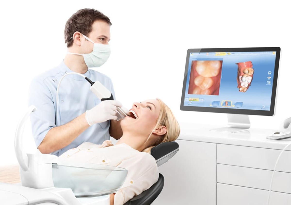
- Improved Patient Education: Intraoral scanners offer the ability to display 3D images of the patient’s oral condition on a screen, allowing dentists to visually explain dental issues and treatment options. This visual representation enhances patient understanding and empowers them to make informed decisions about their oral health.
Overview of Intraoral Scanners
Intraoral scanners have emerged as a game-changing technology in modern dentistry. These compact, handheld devices use cutting-edge technology to capture detailed 3D images of the oral cavity. A closer look at the components and working mechanism of intraoral scanners gives us a better understanding of their capabilities.
An intraoral scanner typically consists of a handheld wand equipped with a small camera and a light projection system. The wand is ergonomically designed to comfortably fit in the dentist’s hand, allowing for easy maneuverability during image acquisition.
The image acquisition process begins by positioning the wand inside the patient’s mouth. The scanner captures multiple images of the teeth, gums, and surrounding structures. These images are then processed in real-time, creating a digital representation of the patient’s oral cavity.
One of the key components of an intraoral scanner is the light projection technology. This technology projects a structured light pattern onto the teeth and gums, allowing the scanner to precisely capture the surface morphology. By analyzing the distortion of the light pattern, the scanner’s software calculates the depth and spatial information to create a highly accurate 3D model.
Intraoral scanners employ different scanning techniques, such as video scanning or still image capturing. Video scanning involves continuously moving the wand throughout the patient’s mouth, capturing a series of frames that are stitched together to create a complete 3D model. On the other hand, still image capturing involves capturing individual images at specific angles and positions to generate the 3D model.
The accuracy and precision of intraoral scanners have significantly improved in recent years. Advanced algorithms and software processing ensure that the digital models accurately represent the patient’s oral structures. This high level of accuracy allows dentists to make more informed clinical decisions and provide optimal treatment outcomes.
Intraoral scanners find extensive application in various dental procedures, including restorative dentistry, orthodontics, implant planning, and smile makeovers. They not only improve the efficiency of these procedures but also enhance patient comfort and satisfaction.
Overall, the advent of intraoral scanners has transformed the way dentistry is practiced. These devices have simplified the imaging process, provided more accurate representations of the oral cavity, and improved treatment planning. As technology continues to advance, we can expect even more sophisticated intraoral scanning systems and further improvements in dental care.
Image Acquisition Process
The image acquisition process is a crucial step in using an intraoral scanner to capture accurate and detailed 3D images of the oral cavity. Let’s explore the stages involved in the image acquisition process and understand how it contributes to the overall scanning procedure.
1. Preparation: Before starting the image acquisition process, the dentist ensures that the patient’s mouth is clean and dry. It is important to remove any debris or fluids that may affect the accuracy of the scan. The dentist may also apply a contrast medium to enhance the visibility of certain structures.
2. Positioning: The dentist positions the handheld wand of the intraoral scanner inside the patient’s mouth. The wand is moved around the different areas of the oral cavity to capture multiple images. The dentist carefully guides the wand to ensure that all the required areas are scanned properly.
3. Image Capture: As the wand is moved, the scanner captures a series of images in real-time. The images are taken from different angles and positions to ensure a complete representation of the oral structures. The camera within the wand collects the visual data required to create the 3D model.
4. Light Projection: The intraoral scanner uses advanced light projection technology to capture the surface morphology of the teeth and gums. It projects a structured light pattern onto the surfaces being scanned. By analyzing the distortion of the light pattern, the scanner’s software calculates the depth and spatial information to create a detailed 3D model.
5. Real-time Processing: The captured images are processed in real-time by the scanner’s software. This processing includes aligning the images, removing any artifacts or noise, and creating a seamless 3D representation. The software ensures that the digital model accurately represents the patient’s oral structures.
6. Verification: Once the image acquisition process is complete, the dentist reviews the generated 3D model on the scanner’s screen or computer monitor. They can navigate through the model, zoom in on specific areas, and verify the accuracy of the scan. This step is crucial to ensure that all the required information has been captured correctly.
The image acquisition process with an intraoral scanner is typically quick and efficient. It eliminates the need for traditional dental impressions, which could take longer and require additional materials. With the advancements in intraoral scanning technology, the process has become more streamlined and comfortable for both the dentist and the patient.
By accurately capturing the oral cavity, the image acquisition process sets the foundation for precise treatment planning and execution. The detailed 3D images obtained through intraoral scanning enable dentists to diagnose dental issues, design customized restorations, and provide optimal oral care to their patients.
Light Projection Technology
Light projection technology is a critical component of intraoral scanners, allowing for the precise capture of the surface morphology of the teeth and gums. By projecting a structured light pattern onto the oral structures, intraoral scanners can gather the necessary information to create accurate 3D models. Let’s dive deeper into how light projection technology works and its significance in the scanning process.
Light projection technology in intraoral scanners involves projecting a pattern of light onto the surfaces being scanned. This pattern can take various forms, such as horizontal or vertical stripes, grids, or random patterns. The purpose of the pattern is to provide reference points for the scanner’s software to calculate the depth and spatial information of the oral structures.
When the light pattern is projected onto the teeth and surrounding tissues, it interacts with the contours and shapes. The reflected or distorted light pattern is captured by the intraoral scanner’s camera, which then processes the information to create a 3D model of the scanned area.
One of the key advantages of light projection technology is its ability to capture the surface details of the oral structures. By analyzing the distortion of the light pattern, the scanner’s software can accurately calculate the position, shape, and texture of the teeth and gums. This level of precision allows for accurate treatment planning and ensures a customized fit for dental restorations.
The accuracy of the light projection technology used in intraoral scanners is continually improving. Advanced algorithms and software enable scanners to capture even the smallest details with high precision. This allows dentists to identify fine cracks, surface irregularities, and other dental conditions that may not be easily visible to the naked eye.
Furthermore, light projection technology plays a crucial role in eliminating potential errors caused by different factors, such as motion or patient movement. The structured light pattern offers a reference point for image alignment, ensuring that the captured images are properly aligned and stitched together to create a seamless 3D model.
Another notable benefit of light projection technology is its ability to work effectively in various lighting conditions. Intraoral scanners are designed to adapt to different lighting environments, ensuring reliable and accurate results regardless of whether the dental procedure is performed in a brightly lit operatory or a more challenging lighting environment.
Intraoral Scanning Techniques
Intraoral scanning techniques are the methods used to capture 3D images of the oral cavity using an intraoral scanner. These techniques vary depending on the scanner’s design and the desired outcome. Let’s explore some common intraoral scanning techniques and their applications in dentistry.
1. Video Scanning: Video scanning involves continuously moving the handheld scanner’s wand throughout the patient’s mouth to capture a series of frames. These frames are then stitched together to create a complete 3D model of the oral structures. Video scanning is commonly used for full-arch scans, capturing the entire upper or lower jaw in a single scan. This technique is useful for planning comprehensive treatment options, such as full-mouth restorations or orthodontic evaluations.
2. Still Image Capturing: Still image capturing involves capturing individual images of specific areas at various angles and positions. The scanner is paused at each position to take a still image before moving on to the next area. These individual images are then aligned and merged to generate a complete 3D model. Still image capturing is commonly used for capturing detailed images of specific teeth or smaller areas of interest. This technique allows for precise evaluations of individual teeth, preparation margins for dental restorations, or localized treatment planning.
3. Single Tooth Scanning: Single tooth scanning focuses on capturing detailed 3D images of a single tooth or a specific area of interest. This technique is beneficial for crown preparations, evaluating specific dental issues, or designing customized dental restorations. By scanning only the necessary area, single tooth scanning reduces scan time and improves efficiency during treatment planning.
4. Bite Registration: In addition to capturing the 3D images of the oral structures, intraoral scanners can also assist in capturing accurate bite registrations. Bite registration involves capturing the way the upper and lower teeth fit together when the patient’s jaws are in their natural position. This information is valuable for diagnosing and planning various dental treatments, such as orthodontic evaluations, occlusal analysis, or designing dental appliances.
5. Scanning with Powder: In instances when the surfaces being scanned lack sufficient contrast or there are reflective surfaces, applying a light-reflective powder can enhance the scanning process. The powder creates a contrasting surface that allows for better detection of details and improved accuracy.
Each scanning technique has its own advantages and applications, and the choice of technique depends on the specific requirements of the dental procedure. Intraoral scanners offer flexibility in capturing scans, enabling dentists to choose the most appropriate technique for accurate diagnosis, treatment planning, and customized solutions.
Accuracy and Precision of Intraoral Scanners
Accuracy and precision are crucial factors to consider when assessing the performance of intraoral scanners. The ability of these advanced imaging devices to capture detailed and reliable 3D images of the oral cavity plays a significant role in treatment planning and execution. Let’s delve into the accuracy and precision of intraoral scanners and their importance in dental practice.
1. Accuracy: Accuracy refers to the degree of conformity between the digital model generated by the intraoral scanner and the actual oral structures. The accuracy of intraoral scanners determines how closely the digital representation matches the real-life anatomy of the patient’s teeth, gums, and surrounding tissues. High accuracy ensures that treatment plans, including the design and fabrication of dental restorations, align with the patient’s specific oral dimensions.
In recent years, intraoral scanners have significantly improved in terms of accuracy. Advancements in hardware and software technologies, along with meticulous calibration procedures, have enhanced the precision of image acquisition. Modern intraoral scanners boast accuracy within microns, allowing for detailed evaluations and precise treatment planning.
2. Precision: Precision refers to the consistency and repeatability of measurements obtained from the intraoral scanner. Precise scanners produce consistent results when capturing multiple scans of the same area. This level of precision ensures that the clinician can confidently rely on the scanner’s measurements for accurate treatment planning and execution.
The precision of intraoral scanners is influenced by various factors such as the scanning technique used, the stability of the scanning device, and the software algorithms employed. Manufacturers place great emphasis on improving the precision of scanners, as it directly impacts the reliability of the digital models generated.
3. Importance in Dental Practice: The accuracy and precision of intraoral scanners have revolutionized dentistry in several ways. These devices provide dentists with the ability to diagnose dental conditions more accurately, plan treatments more precisely, and offer customized solutions tailored to individual patient needs.
By capturing highly accurate 3D images of the oral cavity, intraoral scanners allow for better visualization and evaluation of conditions such as tooth decay, periodontal disease, malocclusions, and other dental pathologies. This precise diagnostic information helps dentists develop appropriate treatment plans and communicate effectively with patients regarding their oral health conditions.
In addition, the accuracy and precision of intraoral scanners improve the fit and aesthetics of dental restorations. Accurate digital models allow for precise design and fabrication of crowns, bridges, aligners, and other dental prosthetics. This not only enhances patient comfort but also ensures optimal long-term outcomes.
The integration of accurate and precise intraoral scanning technology also promotes interdisciplinary collaboration between dental professionals. Digital files generated by intraoral scanners can be easily shared with specialists, dental laboratories, or other healthcare providers, fostering seamless communication and coordinated dental care.
Overall, the accuracy and precision of intraoral scanners are crucial for modern dental practice. These devices have transformed the way dentistry is performed, providing clinicians with invaluable tools for accurate diagnosis, precise treatment planning, and improved patient outcomes.
Applications of Intraoral Scanners in Dentistry
Intraoral scanners have revolutionized the field of dentistry by offering numerous applications across various dental procedures. From diagnosis to treatment planning and fabrication of dental restorations, intraoral scanners have become an invaluable tool in modern dental practice. Let’s explore some of the key applications of intraoral scanners.
1. Restorative Dentistry: Intraoral scanners play a vital role in restorative dentistry. They enable dentists to capture accurate 3D models of the patient’s teeth and gums, allowing for precise planning and fabrication of dental restorations such as crowns, bridges, inlays, and onlays. This technology ensures proper fit, aesthetics, and occlusion, resulting in improved patient satisfaction and long-term restoration success.
2. Orthodontics: Intraoral scanners have transformed the orthodontic workflow by providing digital models that can replace traditional dental impressions for the fabrication of orthodontic appliances, like clear aligners. These scanners streamline the treatment process, allowing for efficient and precise treatment planning, monitoring, and adjustment of orthodontic treatments.
3. Implant Dentistry: In implant dentistry, intraoral scanners are used to capture digital impressions of the implant site and surrounding dentition. These digital impressions enable accurate treatment planning and the fabrication of surgical guides for implant placement. The use of intraoral scanners eliminates the need for messy and uncomfortable conventional impressions, providing a more comfortable experience for patients.
4. Smile Makeovers: Intraoral scanners contribute significantly to smile makeovers and cosmetic dentistry. The digital imaging capabilities of these scanners allow dentists to create virtual smile simulations, enabling patients to see the potential results of their smile transformations before undergoing any treatment. This helps in achieving the desired aesthetic outcomes and ensures patient satisfaction.
5. Periodontics: Intraoral scanners aid in the diagnosis and monitoring of periodontal conditions. The ability of these scanners to capture detailed images of gingival tissues and tooth anatomy allows dentists to assess periodontal health, identify pockets, and plan appropriate periodontal treatments. This technology helps in providing precise treatment and monitoring the progress of periodontal therapy over time.
6. Temporal Monitoring and Comparisons: Intraoral scanners allow dentists to capture digital scans at different time points during the patient’s treatment journey. These sequential scans facilitate comparison and analysis of treatment progress, including assessing changes in tooth movement, occlusal development, and the effectiveness of treatments such as orthodontics or periodontal therapy.
7. Digital Records and Communication: Intraoral scanners generate accurate digital records of patients’ oral conditions, which can be easily stored and accessed for future reference. These digital records also enable seamless communication and collaboration between dentists, specialists, and dental laboratories. Digital files can be shared securely, allowing for efficient treatment planning, case discussion, and coordination of patient care.
The applications of intraoral scanners continue to expand as technology advances. The versatility and precision offered by these devices have transformed various aspects of dentistry, enhancing the accuracy of diagnosis, treatment planning, and the overall quality of dental care.
Comparison of Intraoral Scanners with Traditional Impressions
Intraoral scanners have emerged as an innovative alternative to traditional dental impressions, bringing numerous advantages to both patients and dentists. Let’s compare intraoral scanners with traditional impressions to understand the key differences and benefits of using digital scanning technology.
1. Patient Experience: Traditional dental impressions typically involve placing impression material, such as alginate or silicone, in the patient’s mouth. This can often be uncomfortable and may cause gagging or discomfort. In contrast, intraoral scanners eliminate the need for messy impression materials, providing a more comfortable experience for patients during the scanning process.
2. Time Efficiency: Intraoral scanners offer a significant advantage in terms of time efficiency. Traditional impressions require the setting time for the material before it can be removed from the patient’s mouth. Intraoral scanners, on the other hand, capture digital images in real-time, saving valuable chairside time and reducing overall treatment duration.
3. Accuracy and Precision: Digital scanning technology has significantly improved the accuracy and precision of capturing oral structures compared to traditional impressions. Intraoral scanners can capture highly detailed 3D images, providing dentists with more accurate information for treatment planning and fabrication of dental restorations.
4. Treatment Planning: Intraoral scanners generate digital models that can be easily manipulated and analyzed using computer software. This allows for better treatment planning and customization of dental restorations. Traditional impressions, on the other hand, require physical models that can be more challenging and time-consuming to modify and analyze.
5. Communication and Collaboration: Digital files generated by intraoral scanners can be quickly and securely shared with dental laboratories, specialists, or other dental professionals. This facilitates improved communication and collaboration for treatment planning and execution. Traditional impressions, on the other hand, require physical transportation, which can be time-consuming and may introduce potential errors during the process.
6. Storage and Retrieval: Intraoral scanners create digital records that can be easily stored and accessed for future reference. This eliminates the need for physical storage of traditional impressions, saving space and simplifying record keeping.
7. Cost: While the initial investment in an intraoral scanner may be higher compared to traditional impression materials, the long-term cost benefits can be substantial. Digital scanning eliminates the need for impression materials, trays, and shipping costs associated with traditional impressions. Additionally, fewer retakes or remakes are required with digital scanning, reducing overall costs for both dental professionals and patients.
Overall, intraoral scanners have revolutionized the dental industry by offering a more comfortable, efficient, and accurate alternative to traditional impressions. Their ability to capture detailed 3D digital images enhances treatment planning, improves patient experience, and promotes seamless communication and collaboration within the dental team.
Advantages and Disadvantages of Intraoral Scanners
Intraoral scanners have become an integral part of modern dentistry, offering numerous advantages and transforming the way dental professionals diagnose, plan, and execute treatments. However, like any technology, there are also certain disadvantages to consider. Let’s examine the advantages and disadvantages of using intraoral scanners in dental practice.
Advantages:
1. Improved Patient Experience: Intraoral scanners eliminate the need for messy and uncomfortable traditional impressions, providing a more comfortable experience for patients.
2. Time Efficiency: Intraoral scanners capture digital images in real-time, saving chairside time and reducing overall treatment duration.
3. Accuracy and Precision: Intraoral scanners capture highly detailed 3D images, offering improved accuracy and precision compared to traditional impressions.
4. Treatment Planning: Digital models generated by intraoral scanners allow for better treatment planning, customization of dental restorations, and virtual smile simulations.
5. Communication and Collaboration: Digital files can be easily shared with dental laboratories and specialists, facilitating better communication and collaboration among dental professionals.
6. Storage and Retrieval: Intraoral scanners create digital records that can be easily stored and accessed for future reference, eliminating the need for physical storage of traditional impressions.
7. Cost Efficiency: While the initial investment in an intraoral scanner may be higher, the long-term cost benefits include the elimination of impression materials and shipping costs, as well as reducing retakes or remakes.
Disadvantages:
1. Initial Investment: Intraoral scanners have an upfront cost that may be higher than traditional impression materials.
2. Learning Curve: Dentists and dental staff may require training to effectively operate and integrate intraoral scanners into their workflow.
3. Hardware Limitations: Some intraoral scanners may have limitations in terms of capturing specific areas or angles within the oral cavity.
4. Patient Cooperation: Intraoral scanning requires patient cooperation to ensure proper positioning of the scanner, which may be challenging for some patients.
5. Environmental Factors: Intraoral scanner performance can be affected by factors such as saliva, moisture, or reflective surfaces.
6. Limited Accessibility: In some cases, intraoral scanners may not be accessible or feasible for certain patients or dental practices.
Overall, the advantages of intraoral scanners, including improved patient experience, time efficiency, accuracy, and collaboration, outweigh the disadvantages. With proper training and consideration of limitations, intraoral scanners can significantly enhance dental practice, providing better diagnostic capabilities, treatment planning, and patient satisfaction.
Future Developments in Intraoral Scanning Technology
Intraoral scanning technology has made significant advancements in recent years, revolutionizing the field of dentistry. However, the ongoing development and innovation in this area continue to push the boundaries of what is possible. Let’s explore some exciting future developments and emerging trends in intraoral scanning technology.
1. Improved Accuracy and Precision: Intraoral scanners will likely continue to improve in terms of accuracy and precision. Advancements in hardware and software technologies will allow for even more detailed and accurate digital impressions, enabling dentists to make more precise treatment decisions and fabricate better-fitting dental restorations.
2. Enhanced Speed: Future intraoral scanners are expected to further enhance scanning speed. This will reduce chairside time for patients and increase efficiency in dental practices, enabling dentists to perform more procedures in a shorter amount of time.
3. Artificial Intelligence (AI) Integration: With the integration of AI algorithms, intraoral scanners will become smarter and more intuitive. AI can aid in automatic identification of dental structures, analysis of scans, and assist dentists in treatment planning, improving accuracy, and efficiency.
4. Real-Time Virtual Reality (VR) and Augmented Reality (AR) Visualization: Intraoral scanners may incorporate real-time VR and AR visualization technologies, allowing dentists and patients to view the digital models in an immersive and interactive manner. This can aid in better communication, treatment planning, and patient education.
5. Expanded Applications: Intraoral scanning technology will likely expand its applications beyond restorative dentistry and orthodontics. Emerging applications may include oral pathology assessments, caries detection, and monitoring the progression of dental conditions over time.
6. Smaller, More Ergonomic Designs: Intraoral scanners may become smaller, lighter, and more ergonomic, further improving the operator’s experience and patient comfort during scanning. This can enhance maneuverability and make it easier to capture scans in difficult-to-reach areas of the oral cavity.
7. Integration with Digital Workflows: Future intraoral scanners will seamlessly integrate into digital workflows, allowing for smoother communication and collaboration between dentists, dental laboratories, and other healthcare providers. This integration will facilitate efficient data transfer, streamlined treatment planning, and more coordinated patient care.
8. Enhanced Patient Tracking and Monitoring: Intraoral scanners may incorporate features that enable long-term patient tracking and monitoring of changes in oral health. With regular scanning, dentists can detect early signs of problems, track treatment progress, and identify potential areas of concern to provide proactive and preventive dental care.
As technology continues to rapidly evolve, the possibilities for the future of intraoral scanning technology seem endless. These advancements hold the potential to transform dental practice, making it more efficient, accurate, and patient-centric. Dentists and dental professionals can eagerly anticipate these developments to further enhance the quality of care they provide to patients.