
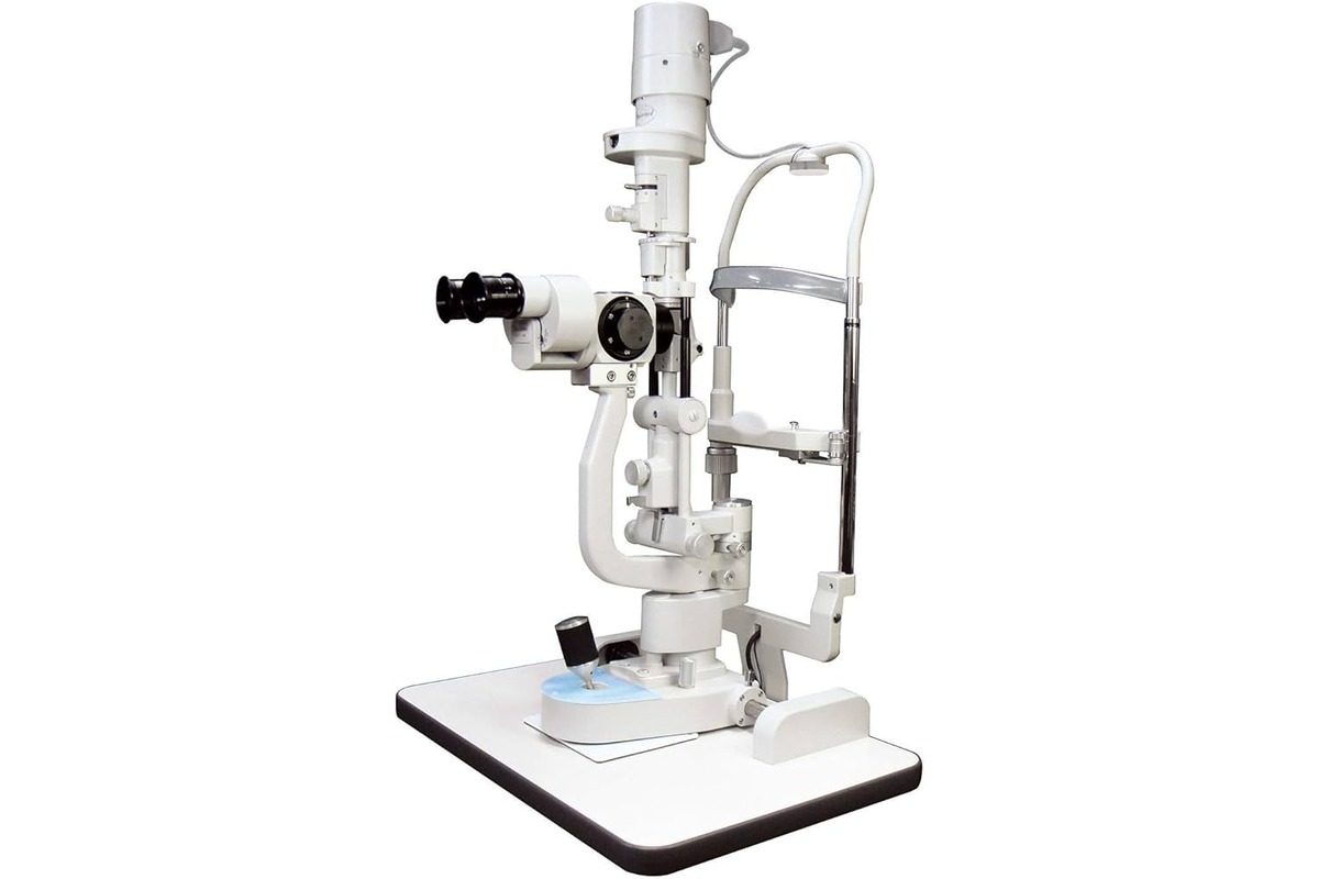
Overview of a Slit Lamp
A slit lamp is a vital instrument used in the field of ophthalmology to examine the structures of the human eye. It consists of a binocular microscope equipped with a high-intensity light source and a slit beam. The slit beam can be adjusted to various widths and angles, allowing for detailed examination of different parts of the eye.
This versatile instrument provides eye care professionals with a magnified view of the anterior segment of the eye, including the cornea, iris, lens, and anterior chamber. It plays a crucial role in diagnosing and monitoring various eye conditions, such as cataracts, glaucoma, and corneal abnormalities.
The slit lamp examination is non-invasive and painless, making it an essential tool in routine eye check-ups and specialized eye care. It allows eye care professionals to evaluate the health of the eye, identify abnormalities, and determine the appropriate treatment plan for each patient.
During the slit lamp examination, patients are seated in front of the instrument and asked to place their chin on a chin rest. The eye care professional then adjusts the microscope and focuses the slit beam on the area of interest. By examining the eye under different lighting conditions and using various techniques, such as retroillumination and sclerotic scatter, the eye care professional can gain valuable insights into the condition of the eye.
With the advancement of technology, modern slit lamps often come with additional features such as filters, cameras, and digital imaging capabilities. These enhancements allow for better visualization, documentation, and sharing of eye examination results, facilitating collaboration between eye care professionals and enhancing patient care.
Components of a Slit Lamp
A slit lamp consists of several essential components that work together to enable detailed examination of the eye:
- Microscope: The microscope of a slit lamp provides high magnification and clarity, allowing the eye care professional to view the eye structures in detail. It typically has binocular eyepieces, which provide a three-dimensional view, enhancing depth perception.
- Slit Beam: The slit beam is a thin, adjustable light source that is projected onto the eye. It can be customized in terms of width, intensity, and angle, providing different viewing angles and illumination techniques for specific examinations.
- Chin Rest: The chin rest helps stabilize the patient’s head and ensures that they maintain a steady position during the examination. This allows for accurate and consistent images to be obtained.
- Joystick or Control Handles: These controls allow the eye care professional to adjust the focus, aperture, and angle of the slit beam. They also enable movement of the microscope to examine different areas of the eye.
- Filters: Slit lamps often have various filters that can be interchanged to enhance specific aspects of the examination. For example, a blue filter can help detect corneal abrasions, while a red-free filter can highlight blood vessels in the eye.
- Illumination Source: Slit lamps use a high-intensity light source, such as a halogen or LED bulb, to provide consistent and bright illumination during the examination.
- Beam Splitter: Some advanced slit lamps are equipped with a beam splitter, which allows simultaneous viewing through the microscope eyepieces and recording the examination using a camera or digital imaging system.
- Apertures: Slit lamps often have interchangeable apertures that can be used to modify the shape and size of the slit beam. Different apertures are selected based on the specific requirements of the examination.
Each component of a slit lamp is meticulously designed to provide optimal visualization and examination capabilities. The combination of these components allows eye care professionals to accurately diagnose and monitor various eye conditions, ensuring the delivery of high-quality eye care.
Importance of a Slit Lamp in Ophthalmology
The slit lamp is an indispensable tool in the field of ophthalmology, playing a crucial role in the evaluation and management of eye conditions. Here are some key reasons why the slit lamp is of paramount importance:
1. Detailed Examination: The slit lamp enables eye care professionals to perform a thorough examination of the anterior segment of the eye. This includes assessing the cornea, iris, lens, and anterior chamber, allowing for the detection of abnormalities or diseases that might not be visible through other examination methods.
2. Precise Diagnosis: The high magnification and illumination provided by the slit lamp aid in the accurate diagnosis of various eye conditions. Eye care professionals can visualize and evaluate minute details, such as corneal abrasions, foreign bodies, or signs of infection, helping them make informed diagnostic decisions.
3. Monitoring Progression: The slit lamp is invaluable in monitoring the progression of eye conditions over time. By performing regular slit lamp examinations, eye care professionals can assess changes in the eye structures, measure the effectiveness of treatments, and make necessary adjustments to the patient’s management plan.
4. Treatment Planning: The detailed view provided by the slit lamp assists in planning appropriate treatment strategies for patients. It helps determine the best course of action, whether it’s prescribing medications, recommending surgical intervention, or referring the patient to a specialist for further evaluation.
5. Patient Education: The slit lamp examination provides an opportunity for eye care professionals to educate patients about their eye health. By visually demonstrating the condition of their eyes and explaining the significance of certain findings, patients gain a better understanding of their eye condition and the importance of adhering to treatment plans.
6. Research and Teaching: Slit lamps are extensively used in ophthalmic research and educational settings. They allow researchers to study eye structures and pathologies in detail, contributing to advancements in the field. Additionally, slit lamp examinations are an essential part of ophthalmology training, enabling medical students and residents to develop their skills in eye examination and diagnosis.
Overall, the slit lamp is a cornerstone of ophthalmic practice, providing eye care professionals with invaluable information to diagnose, monitor, and manage a wide range of eye conditions. Its importance in delivering comprehensive patient care cannot be overstated, making it an indispensable tool in the field of ophthalmology.
How Does a Slit Lamp Work?
A slit lamp is a sophisticated instrument that utilizes a combination of illumination and magnification to examine the anterior segment of the eye. Understanding how a slit lamp works is essential in utilizing its capabilities effectively. Here’s a breakdown of its workings:
1. Illumination: The slit lamp uses a high-intensity light source, such as a halogen or LED bulb, to produce a bright and focused beam of light. This light is directed onto the eye structures, illuminating them for examination.
2. Slit Beam: The light emitted from the slit lamp is filtered through an adjustable slit aperture, resulting in a thin, vertical slit beam. The slit beam can be modified in terms of width, angle, and intensity to suit the specific needs of the examination.
3. Microscope System: The slit lamp incorporates a binocular microscope, equipped with eyepieces, objective lenses, and a mechanism for adjusting the focus. This microscope system provides a high-resolution, magnified view of the eye structures.
4. Patient Positioning: The patient is positioned in front of the slit lamp, resting their chin on a chin rest and forehead against a headrest. This ensures stability and proper alignment of the eye for examination.
5. Viewing Technique: The eye care professional uses the binocular eyepieces to view the eye structures through the microscope. The slit beam is then directed onto the area of interest in the anterior segment of the eye.
6. Observation Techniques: Different observation techniques can be employed during slit lamp examination, depending on the specific purpose. These include direct observation, retroillumination (illumination from behind the structures), sclerotic scatter (illumination of the sclera), and specular reflection techniques.
7. Filters and Accessories: Slit lamps often have interchangeable filters and accessories to enhance the examination. Filters can be used to emphasize or suppress certain wavelengths of light for better visualization of specific structures or conditions.
8. Documentation and Recording: Advanced slit lamps may include a beam splitter mechanism that allows simultaneous viewing through the eyepieces and capturing images or videos of the examination. This enables the documentation and analysis of the findings for further evaluation or patient records.
By combining precise illumination and magnification, a slit lamp provides eye care professionals with a detailed and enhanced view of the anterior segment of the eye. It allows for the identification of subtle abnormalities, precise measurement of structures, and accurate diagnosis of various eye conditions.
Advantages of a Slit Lamp in Eye Examination
The slit lamp is widely recognized as an invaluable instrument in eye examinations. It offers numerous advantages that contribute to accurate diagnosis, efficient treatment, and improved patient care. Here are some key advantages of using a slit lamp:
1. Enhanced Visualization: The slit lamp provides a highly magnified view of the anterior segment of the eye, allowing eye care professionals to visualize even the tiniest details of the eye structures. This enhanced visualization enables the detection of subtle abnormalities or conditions that may not be visible with the naked eye.
2. Precise Localization: With the adjustable slit beam, the eye care professional can precisely localize and examine specific areas of the eye. By controlling the width and angle of the slit beam, they can focus on the cornea, lens, iris, or other structures, facilitating accurate assessment and diagnosis.
3. Comprehensive Examination: The slit lamp enables a comprehensive examination of the anterior segment, including the cornea, conjunctiva, anterior chamber, iris, and lens. It allows for the evaluation of various eye conditions, such as corneal abrasions, cataracts, conjunctivitis, and foreign bodies.
4. Illumination Control: The slit lamp provides control over the intensity, angle, and width of the slit beam, allowing for optimal illumination of the eye structures. This control helps in highlighting specific features, assessing depth, and enhancing visualization of abnormalities or pathologies.
5. Non-invasive and Painless: Slit lamp examinations are non-invasive and painless, making them comfortable for patients. By simply sitting in front of the instrument and resting their chin on the chin rest, patients can undergo a thorough examination without any discomfort or need for anesthesia.
6. High-Quality Imaging: Modern slit lamps often come equipped with digital imaging capabilities, allowing for high-quality imaging and documentation of the examination findings. This not only assists in accurate diagnosis but also provides a visual reference for tracking changes over time and facilitating collaboration between eye care professionals.
7. Versatility and Adaptability: Slit lamps can be customized with various filters, lenses, and accessories to adapt to different examination techniques and specific patient needs. This versatility allows for a wide range of examinations and enhances the overall flexibility of the instrument.
8. Teaching and Research: Slit lamps are valuable tools in teaching settings and research labs. They enable students and researchers to learn and study eye structures, pathologies, and treatment modalities, contributing to the advancement of ophthalmic knowledge and improving patient care.
The advantages offered by a slit lamp make it an indispensable instrument in eye examinations. Its ability to provide detailed visualization, accurate diagnosis, and efficient documentation ensures that eye care professionals can deliver the highest standard of care to their patients.
Different Types of Slit Lamps
Slit lamps come in various types and designs, each catering to specific needs and preferences in eye examinations. Here are the different types of slit lamps commonly used in ophthalmology:
1. Traditional Slit Lamps: Traditional slit lamps consist of a binocular microscope mounted on a stand. They typically feature a single light source and manual adjustment of the slit beam, aperture, and magnification. These slit lamps are widely used in clinics and hospitals for routine eye examinations and basic procedures.
2. Digital Slit Lamps: Digital slit lamps are equipped with advanced imaging and documentation capabilities. They incorporate high-resolution digital cameras that allow for capturing images and videos of the examination. These images can be stored, analyzed, and shared electronically, enabling more efficient communication between eye care professionals and enhancing patient care.
3. Handheld Slit Lamps: Handheld slit lamps are portable and battery-operated, making them convenient for use in remote or mobile clinics and during eye examinations in non-traditional settings. These slit lamps provide flexibility and ease of use, enabling eye care professionals to perform examinations in various environments.
4. Zoom Slit Lamps: Zoom slit lamps feature a variable magnification system, allowing for adjustable levels of magnification during the examination. This enables eye care professionals to switch between different levels of magnification without the need to change the objective lenses. It provides enhanced flexibility and convenience in detailed observation and examination.
5. Modular Slit Lamps: Modular slit lamps offer versatility by allowing for the integration of additional features and accessories. These include filters, imaging modules, and tonometry capabilities. The modular design allows eye care professionals to customize the slit lamp based on their specific needs, expanding its functionality and adaptability.
6. LED Slit Lamps: LED slit lamps utilize energy-efficient LED light sources as the illumination system. LED lights offer long product life, consistent brightness, and reduced heat emission. These slit lamps are environmentally friendly and provide a stable light source, ensuring reliable and accurate eye examinations.
7. Infrared Slit Lamps: Infrared slit lamps utilize infrared light instead of visible light for examination purposes. This enables eye care professionals to visualize structures that might not be visible under normal illumination conditions, such as the angle of the eye or corneal inflammation.
Each type of slit lamp offers unique features and benefits, catering to the diverse needs of eye care professionals. The choice of the slit lamp depends on the specific requirements of the examination and the preferences of the ophthalmologist or optometrist.
How to Use a Slit Lamp Correctly
Using a slit lamp correctly is essential to ensure accurate examination and diagnosis. Here are some important steps to follow when using a slit lamp:
1. Preparation: Before beginning the examination, ensure that the slit lamp is properly calibrated and functioning correctly. Adjust the height, tilt, and angle of the microscope to accommodate patient comfort and proper alignment with the eye.
2. Patient Positioning: Instruct the patient to sit comfortably and place their chin on the chin rest. Adjust the height and position of the chin rest to ensure proper alignment of the patient’s eye with the microscope. Make sure the patient’s forehead is resting against the headrest for stability.
3. Eye Care Professional Positioning: Position yourself comfortably behind the microscope, aligning your eyes with the eyepieces. Adjust the focus and interpupillary distance of the microscope to ensure a clear view of the patient’s eye.
4. Illumination and Slit Beam: Adjust the illumination settings based on the desired examination technique. Control the width, angle, and intensity of the slit beam to illuminate the specific area of interest in the eye. Use filters if necessary to enhance visualization of certain structures or conditions.
5. Visual Inspection: Begin the examination by visually inspecting the eye structures under low magnification. Observe the eyelids, lash line, tear film, conjunctiva, and sclera for any abnormalities or signs of inflammation.
6. Slit Beam Techniques: Employ various slit beam techniques to examine different structures and conditions. These techniques include direct observation, retroillumination, sclerotic scatter, and specular reflection. Modify the slit beam width and angle to focus on specific areas and achieve optimal visualization.
7. Magnification and Fine Focus: Gradually increase the magnification to perform a detailed examination of the cornea, iris, lens, and anterior chamber. Use fine focus adjustments to achieve clear and sharp images.
8. Documenting Findings: If equipped, use the camera or imaging system to capture images or record videos of the examination findings. Properly label and save the documentation for future reference, patient records, or consultation purposes.
9. Patient Communication: Throughout the examination, communicate with the patient, explaining the procedure, findings, and any recommended actions or treatments. Encourage the patient to ask questions and address any concerns they may have.
10. Cleanliness and Maintenance: After completing the examination, ensure that the slit lamp is properly sanitized and cleaned according to the manufacturer’s instructions. Regularly maintain and calibrate the instrument to ensure consistent and accurate performance.
By following these guidelines, eye care professionals can effectively and correctly use a slit lamp to conduct thorough and accurate examinations, enabling precise diagnosis and appropriate management of various eye conditions.
Common Applications of a Slit Lamp in Eye Care
The slit lamp is a versatile instrument that finds wide application in various aspects of eye care. Here are some common applications of the slit lamp in diagnosing and managing eye conditions:
1. Corneal Examination: The slit lamp allows for detailed examination of the cornea, including assessing its transparency, detecting irregularities, evaluating the tear film, and identifying corneal abrasions or ulcers.
2. Anterior Chamber Evaluation: The anterior chamber, which includes the iris, lens, and the space between them, can be thoroughly examined using the slit lamp. This evaluation helps in diagnosing conditions such as anterior uveitis, cataracts, and lens abnormalities.
3. Gonioscopy: Gonioscopy involves the use of special contact lenses and the slit lamp to evaluate the angle of the eye, where the iris meets the cornea. It aids in the diagnosis and management of glaucoma and other conditions related to the drainage of aqueous humor.
4. Contact Lens Assessment: Slit lamps are used to assess the fit, positioning, and condition of contact lenses. This evaluation is essential to ensure the comfort and optimal vision of patients who use contact lenses for correction or therapeutic purposes.
5. Evaluation of Conjunctiva and Sclera: The slit lamp enables the examination of the conjunctiva and sclera, detecting conditions such as conjunctivitis, pterygium, scleritis, and other inflammatory or infectious conditions affecting these tissues.
6. Evaluation of the Iris and Pupils: By adjusting the slit beam, eye care professionals can assess the iris for abnormalities, such as iris atrophy, nevi, or iris dilator muscle function. The size and reaction of the pupils can also be evaluated using the slit lamp, aiding in the diagnosis and management of conditions affecting pupillary responses.
7. Examination of the Lens and Posterior Segment: Although the slit lamp primarily focuses on the anterior segment, it can provide limited visualization of the lens and posterior segment structures. This aids in the assessment of conditions such as posterior subcapsular cataracts and allows for the detection of any vitreous abnormalities.
8. Foreign Body Detection and Removal: Slit lamps are useful in locating and removing foreign bodies from the cornea or conjunctiva. The focused illumination and magnification provided by the slit lamp enable eye care professionals to safely and accurately remove these objects without causing further damage.
9. Monitoring Eye Conditions: Slit lamps are essential in monitoring the progression and response to treatment of various eye conditions. Regular slit lamp examinations help eye care professionals track changes in the eye structures and make informed decisions regarding treatment adjustment or intervention.
10. Patient Education and Counseling: Slit lamp examinations provide eye care professionals with visual aids to explain and educate patients about their eye health and conditions. By using the slit lamp findings, patients can better understand their eye condition and actively participate in their treatment plan.
The applications of a slit lamp in eye care span a wide range of examinations, allowing for accurate diagnosis, monitoring of conditions, and effective management of eye health.
Tips for Maintaining and Cleaning a Slit Lamp
Maintaining and cleaning a slit lamp is crucial for its proper functioning, longevity, and accuracy of examination. Here are some important tips to follow for maintaining and cleaning a slit lamp:
1. Regular Cleaning: Clean the slit lamp after each use to remove any dust, debris, or remnants of ocular fluids. Use a lint-free cloth or a soft brush to gently wipe the surfaces of the microscope, chin rest, and other exposed parts.
2. Sanitization: Practice proper sanitization procedures to prevent the spread of infections. Use an approved disinfectant solution or wipes to sterilize the surfaces that come into contact with the patient, such as the chin rest and forehead rest.
3. Follow Manufacturer’s Instructions: Always refer to the manufacturer’s instructions and guidelines for specific cleaning and maintenance recommendations for your slit lamp model. Different brands and models may have unique requirements.
4. Calibration and Maintenance Checks: Regularly schedule professional calibration and maintenance checks for your slit lamp. This ensures accurate alignment, proper functioning of the illumination system, and optimization of the imaging and measurement components.
5. Replacement of Filters and Bulbs: Replace worn-out or expired filters and bulbs according to the manufacturer’s recommendations. This ensures consistent and accurate illumination during examinations.
6. Lubrication of Moving Parts: If applicable, periodically apply lubricant to moving parts of the slit lamp to ensure smooth and precise adjustments. Refer to the manufacturer’s guidelines to identify the appropriate lubricant and frequency of application.
7. Protection from Environmental Factors: Keep the slit lamp protected from extreme temperatures, humidity, and direct sunlight, as these can affect its performance and longevity. Store it in a clean, dry, and controlled environment when not in use.
8. Proper Handling and Transport: Handle the slit lamp with care, avoiding any rough or improper handling that may cause damage. During transport, secure the instrument to prevent any jarring movements or vibrations that may affect its components.
9. Maintain a Clean Environment: Ensure the examination room or clinic is kept clean and free from dust, moisture, or other environmental contaminants that may affect the slit lamp’s performance. Regularly clean the area surrounding the slit lamp to prevent debris from getting onto the microscope or other sensitive parts.
10. Staff Training: Provide proper training and education to staff members responsible for using and maintaining the slit lamp. This includes understanding cleaning protocols, recognizing signs of malfunction, and knowing when to seek professional assistance for repairs or maintenance.
By following these tips, eye care professionals can ensure that their slit lamp remains clean, accurate, and in optimal condition for reliable examination and diagnosis. Proper maintenance and cleaning contribute to the overall efficiency and longevity of the instrument, enhancing the quality of patient care.
Potential Risks and Safety Precautions while Using a Slit Lamp
While using a slit lamp in eye examinations, it is crucial to be aware of potential risks and to follow safety precautions to ensure the well-being of both the patient and the eye care professional. Here are some common risks and safety precautions to consider:
1. Eye Exposure to High-Intensity Light: The intense light emitted from the slit lamp can be potentially harmful to the eyes. Eye care professionals should always avoid looking directly into the beam and instruct the patient to keep their eyes closed when adjusting the slit beam or switching between examination techniques.
2. Contamination and Infections: Slit lamps can become contaminated with infectious agents if proper cleaning and sanitization protocols are not followed. Eye care professionals should ensure that all surfaces that come into contact with the patient are sanitized before and after each use to minimize the risk of cross-contamination.
3. Patient Comfort and Safety: It is essential to position the patient correctly and provide adequate support and stability while using the slit lamp. Ensure that the chin rest and forehead rest are adjusted appropriately to prevent discomfort or strain for the patient during the examination.
4. Foreign Body Risk: During examination or while performing procedures, small foreign bodies can inadvertently enter the patient’s eye. Eye care professionals should take precautions to minimize this risk, such as instructing the patient to avoid touching or rubbing their eyes and using a protective shield or lubricating eye drops when necessary.
5. Electrical Safety: Slit lamps require electricity to function, and faulty wiring or improper handling can lead to electrical hazards. Eye care professionals should make sure that the instrument is plugged into a grounded electrical outlet, follow proper electrical safety protocols, and handle the instrument with dry hands.
6. Sudden Movements: The slit lamp and its components should be handled with care to avoid sudden movements or jarring that may cause injury to the patient. Proper positioning, stability, and controlled adjustments should be maintained throughout the examination process.
7. Proper Ventilation: Slit lamps often have illumination systems that generate heat. Eye care professionals should ensure that the instrument is placed in a well-ventilated area to prevent overheating and maintain proper functioning.
8. Maintenance and Calibration: Regular maintenance and calibration are essential to ensure the accuracy and safety of slit lamps. Eye care professionals should follow the manufacturer’s recommendations for maintenance and arrange professional servicing when necessary.
9. Staff Education and Training: It is crucial to provide adequate training to staff members who operate the slit lamp, ensuring they are knowledgeable about safety precautions, proper handling techniques, and protocols for cleaning and sanitization. Continual education and training help maintain a safe working environment.
10. Compliance with Regulatory Standards: Eye care professionals should adhere to local regulations and guidelines related to the use, maintenance, and safety of slit lamps. This includes following infection control protocols, ensuring staff competency, and maintaining proper documentation of equipment maintenance.
By being aware of the potential risks and implementing appropriate safety precautions, eye care professionals can create a safe environment for both themselves and their patients during slit lamp examinations. Prioritizing safety reduces the risk of accidents, improves patient satisfaction, and upholds professional standards of care.