
How Does a Retinal Scanner Work?
A retinal scanner is a sophisticated biometric technology that employs advanced imaging techniques to capture unique patterns in the human retina for identification and authentication purposes. This cutting-edge technology operates on the principle that every person’s retina has distinct characteristics that remain unchanged throughout their lifetime.
The process of retinal scanning involves the use of near-infrared light to illuminate the eye, while a specialized camera captures detailed images of the unique blood vessels and patterns on the retina. The captured images are then converted into a digital format for analysis and comparison.
Retinal scanners primarily rely on the concept of vascular pattern recognition. The blood vessels in the retina form intricate patterns that are unique to each individual, similar to a fingerprint. These patterns can be compared to a pre-stored database of retinal images to determine the person’s identity or authenticate their access to a secured area.
The technology behind retinal scanning is highly precise and accurate. It can identify an individual with an incredibly low margin of error, making it one of the most reliable biometric identification techniques available today.
When a person undergoes a retinal scan, the scanner emits a safe, low-intensity infrared light towards the eye. The light penetrates the various layers of the eye and is absorbed by the blood vessels in the retina. The reflected or scattered light is then captured by the camera, producing a high-resolution image.
One of the key advantages of retinal scanning is its immunity to external factors that may affect other biometric technologies. The patterns in the retina are stable and do not change significantly over time, ensuring consistent and accurate identification. Moreover, the uniqueness of the retinal patterns makes it virtually impossible for someone to replicate or forge another person’s retina.
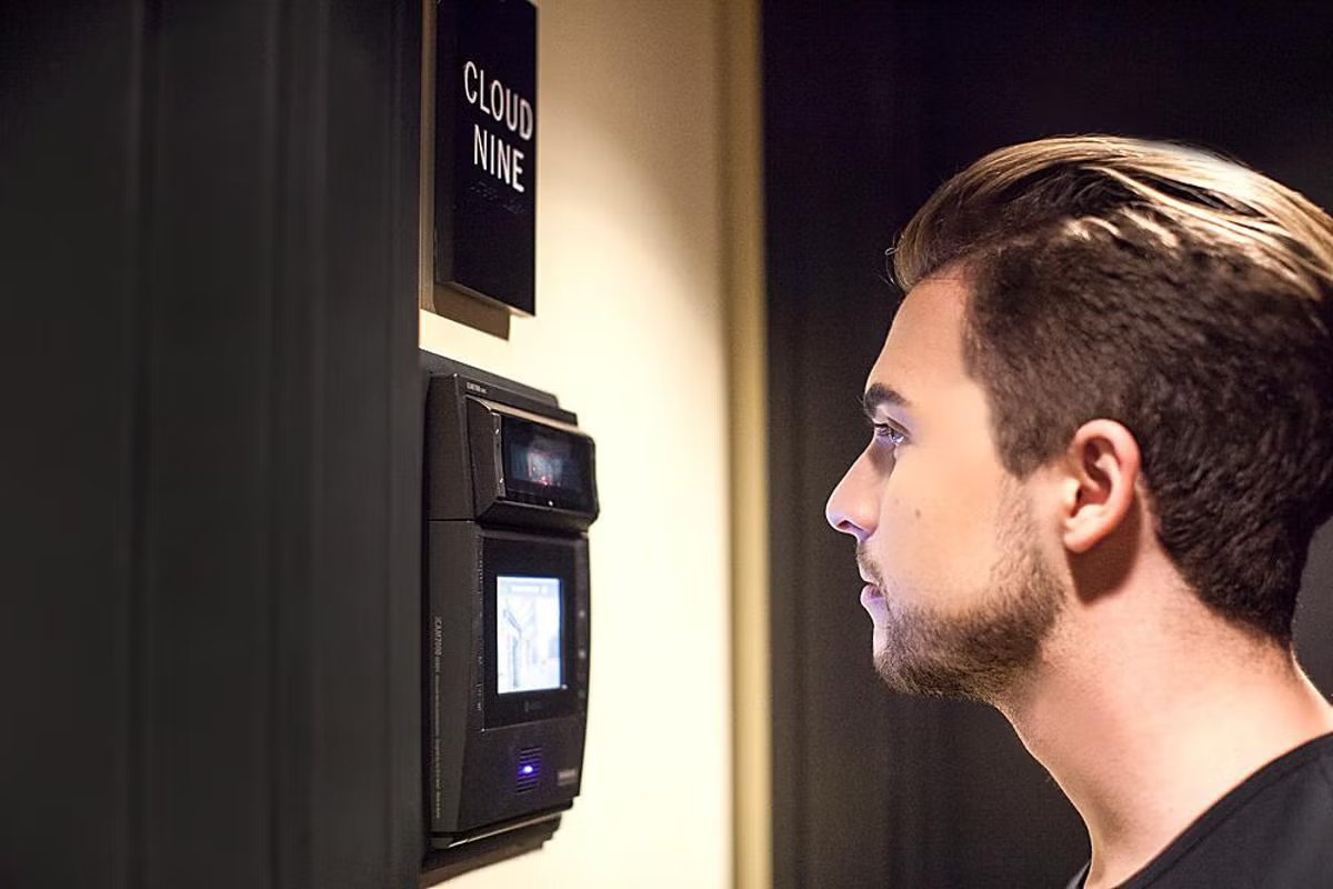
However, the process of retinal scanning also has its limitations and concerns. To undergo a scan, users must position their eyes close to the scanning device, which can impose discomfort for some individuals. Additionally, retinal scanners tend to be expensive and require specialized equipment, limiting their widespread implementation.
Despite these limitations, retinal scanning has found applications in various industries, including access control systems, high-security facilities, banking, healthcare, and government agencies. Its unparalleled accuracy and uniqueness make it an invaluable tool for ensuring secure identification and preventing unauthorized access.
What is a Retinal Scanner?
A retinal scanner is a sophisticated biometric device that utilizes advanced technology to capture and analyze the unique patterns of blood vessels in the retina. The retina, located at the back of the eye, consists of a complex network of blood vessels that supply oxygen and nutrients to the surrounding tissues.
A retinal scanner works by emitting a low-intensity beam of near-infrared light towards the eye. This light is absorbed by the blood vessels in the retina, while the surrounding tissues reflect it back towards the scanner. The scanner then captures the reflected light and converts it into a digital image that represents the intricate patterns of the retina.
The captured digital image is then analyzed using sophisticated algorithms to identify and authenticate individuals. Each person’s retina has unique characteristics that remain stable throughout their life, similar to a fingerprint. These characteristics include the arrangement of blood vessels, their size and shape, and the spacing between them. By analyzing these unique patterns, a retinal scanner can accurately identify individuals with a high level of precision.
Retinal scanners are designed to provide secure and reliable identification and authentication. Compared to other biometric technologies, such as fingerprint or facial recognition, retinal scanning offers a higher level of accuracy. The intricate and complex patterns of the retina make it extremely difficult for someone to duplicate or forge another person’s retina, enhancing the security of the system.
It is important to note that retinal scanners do not pose any health risks to the individual being scanned. The low-intensity near-infrared light used by the scanner is safe and does not cause any harm to the eyes. However, individuals with certain eye conditions, such as cataracts or severe astigmatism, may not be suitable candidates for retinal scanning.
Retinal scanners have found applications in various industries and sectors. They are commonly used in high-security facilities, such as government buildings, military installations, and research laboratories, to control access and ensure only authorized individuals are granted entry. Retinal scanning is also employed in the healthcare industry for patient identification, ensuring accurate medical records and preventing identification errors.
The Anatomy of the Eye
The eye is a remarkable organ that allows us to see and perceive the world around us. It consists of several intricate components working together to capture, focus, and transmit visual information to the brain. Understanding the anatomy of the eye is crucial to comprehend how a retinal scanner works.
The eye can be broadly divided into three main layers: the outer layer, middle layer, and inner layer.
The outer layer of the eye consists of the cornea and the sclera. The transparent cornea is the front part of the eye that covers the iris and the pupil, and it plays a vital role in focusing incoming light. The sclera, commonly known as the white of the eye, is the tough, protective outer covering that maintains the shape of the eyeball.
The middle layer of the eye is called the uvea and contains three main structures: the iris, the ciliary body, and the choroid. The iris is the colored part of the eye that surrounds the pupil and controls the amount of light entering the eye by adjusting the size of the pupil. The ciliary body is responsible for producing the clear fluid called aqueous humor, which nourishes the eye and maintains its shape. The choroid is a highly vascular layer that provides oxygen and nutrients to the retina.
The inner layer of the eye is the retina, which is located at the back of the eye. The retina contains millions of specialized cells called photoreceptors – namely cones and rods – that convert incoming light into electrical signals. These signals are then transmitted through the optic nerve to the brain, where they are processed and interpreted as visual information.
The retina itself consists of several layers, including the ganglion cell layer, bipolar cell layer, and the photoreceptor layer. The photoreceptor layer contains the rods and cones that are responsible for capturing light and transmitting the signals to the brain. The rods are more sensitive to dim light, while the cones are responsible for color vision and function best in bright light conditions.
At the center of the retina lies a small area called the macula, which contains a high concentration of cones and is responsible for producing sharp central vision. Within the macula, there is a small depression called the fovea, which is responsible for the sharpest and most detailed vision.
Understanding the anatomy of the eye and the role of the retina is essential for comprehending how a retinal scanner captures and analyzes the unique patterns in the blood vessels of the retina. By harnessing the intricacies of the eye, retinal scanners provide a secure and reliable method of biometric identification and authentication.
The Retina: An Overview
The retina is a vital component of the eye that plays a crucial role in our ability to see and perceive visual information. Located at the back of the eye, the retina is a complex layer of tissue consisting of millions of specialized cells known as photoreceptors.
The main function of the retina is to convert incoming light into electrical signals that can be interpreted by the brain as visual images. It accomplishes this through the intricate interactions of various cell types within its layers.
There are two main types of photoreceptors in the retina: cones and rods. Cones are responsible for color vision and function best in bright light conditions. They are concentrated mainly in the central part of the retina, called the macula, and particularly in a small area within the macula known as the fovea. The fovea is responsible for providing the sharpest and most detailed vision.
Rods, on the other hand, are more sensitive to dim light and are responsible for peripheral and night vision. They are spread throughout the entire retina, except for the fovea. Rods provide us with the ability to see objects in low-light environments, although they do not perceive color as effectively as cones.
When light enters the eye, it passes through the various layers of the retina before reaching the photoreceptor layer. The photoreceptors contain light-sensitive pigments that undergo chemical changes upon exposure to light. These changes trigger a cascade of electrical reactions within the photoreceptor cells, resulting in the generation of electrical signals.
After the photoreceptor cells capture and convert light into electrical signals, these signals are processed and transmitted to other retinal cells, such as bipolar cells and ganglion cells. Bipolar cells receive signals from the photoreceptors and transmit them to ganglion cells, which act as the final output neurons of the retina.
The axons of ganglion cells gather at the optic disc, a small area on the retina where the optic nerve exits the eye. The optic nerve carries the electrical signals from the retina to the brain, specifically to the visual cortex, where they are further processed and interpreted as visual images.
Understanding the structure and function of the retina is essential in appreciating how a retinal scanner captures and analyzes the unique patterns of blood vessels in the retina. By harnessing the intricate network of cells within the retina, retinal scanners provide a highly accurate and reliable method of biometric identification and authentication.
How Does a Retinal Scanner Capture Images?
A retinal scanner utilizes advanced imaging techniques to capture detailed images of the retina, which are then used for identification and authentication purposes. The process involves several steps that work together to ensure accurate and reliable image capture.
When a person undergoes a retinal scan, they are positioned in front of the scanning device, usually with their forehead against a support and instructed to focus on a specific point. The scanner emits a low-intensity near-infrared light towards the eye, which is safe and does not cause any discomfort or harm.
The near-infrared light is directed towards the retina, where it is absorbed by the blood vessels. The blood vessels in the retina act as natural contrast agents that help distinguish the unique patterns and structures within the retina. Meanwhile, the surrounding tissues reflect or scatter the light back towards the scanner, which captures the reflected light using a specialized camera.
The camera in the retinal scanner is equipped with filters that block out other wavelengths of light, allowing only the near-infrared light to be captured. This helps enhance the clarity and accuracy of the retinal image.
The camera records the reflected light and converts it into a digital image, creating a high-resolution representation of the intricate blood vessel patterns in the retina. The captured image is then processed and stored in a database for future comparison and identification purposes.
The captured image of the retina consists of millions of data points, representing the veins, arteries, and capillaries within the retina. These unique patterns are highly distinctive and vary from individual to individual, similar to a fingerprint.
The retinal image that is captured undergoes various preprocessing steps to enhance the quality and clarity of the image. This may include noise reduction, contrast adjustment, and image normalization techniques to ensure consistent and accurate comparison with stored images in the database.
It is important to note that retinal scanning is a non-intrusive process that does not require physical contact with the eye. The low-intensity near-infrared light used in the scanning process is safe and poses no health risks to the individual being scanned.
Overall, the process of capturing retinal images using a retinal scanner involves the emission of near-infrared light towards the retina, the reflection and scattering of light by the blood vessels and surrounding tissues, and the subsequent capture and conversion of the reflected light into a digital image. This advanced imaging technique enables retinal scanners to provide accurate and reliable biometric identification and authentication.
The Process of Iris Recognition
Iris recognition is a biometric technology that utilizes the unique patterns in the iris of an individual’s eye for identification and authentication purposes. The iris, which is the colored part of the eye surrounding the pupil, contains intricate patterns that are highly distinctive and remain stable throughout a person’s life.
The process of iris recognition involves several steps that work together to capture, analyze, and compare the iris patterns. Let’s explore each step in detail:
1. Image Capture: The first step in iris recognition is to capture a high-resolution image of the iris. This is done using a specialized camera that is designed to specifically focus on the iris. The individual being scanned is asked to position their eyes correctly in front of the camera, and multiple images are captured to ensure clarity and accuracy.
2. Preprocessing: The captured iris image undergoes preprocessing to enhance its quality and remove any noise or artifacts. This may involve techniques such as image cropping, noise reduction, and contrast enhancement to ensure a clear and focused image of the iris.
3. Feature Extraction: In this step, unique features and patterns are extracted from the iris image. The iris is analyzed for characteristics such as furrows, striations, and crypts. These features are then encoded into a mathematical representation known as an iris code. The iris code contains the essential information necessary for comparison and identification.
4. Template Creation: The iris code is used to create a template, which is a compact representation of the iris features. The template serves as a reference for future comparisons and is securely stored in a database.
5. Matching: When an individual presents their iris for recognition, their iris image is captured and processed in the same way as during enrollment. The extracted features are compared with the stored template using complex algorithms to determine if there is a match. The level of similarity between the extracted features and the template is measured using a score or similarity metric.
6. Decision Making: Based on the similarity score, a decision is made regarding the identification or authentication of the individual. If the similarity score exceeds a predefined threshold, the individual is considered a match, and their identity is confirmed. If the similarity score falls below the threshold, the individual is not recognized, and their access may be denied.
Iris recognition offers several advantages as a biometric technology. The iris is highly distinctive, making it difficult for someone to replicate or forge another person’s iris patterns. Additionally, iris recognition is highly accurate, even in conditions where other biometric technologies may struggle, such as low lighting or with individuals wearing glasses or contact lenses.
Overall, the process of iris recognition involves capturing a high-resolution image of the iris, preprocessing and extracting unique features, creating a template for comparison, matching the extracted features with the stored template, and making a decision based on the similarity score. This advanced technology allows for secure and reliable identification and authentication of individuals based on their unique iris patterns.
The Science Behind Retinal Scan Recognition
Retinal scan recognition is based on the intricate science of analyzing and comparing the unique patterns of blood vessels in the retina. The blood vessels in the retina form complex and distinctive patterns that are unique to each individual. Understanding the science behind retinal scan recognition helps to appreciate the precision and reliability of this biometric technology.
One of the key components of retinal scan recognition is vascular pattern recognition. The vasculature of the retina consists of a network of veins, arteries, and capillaries that supply oxygen and nutrients to the retinal tissue. These vessels have distinct characteristics such as size, shape, branching patterns, and spacing.
The first step in retinal scan recognition is capturing a high-resolution image of the retina using a specialized camera. This camera uses near-infrared light, which can easily penetrate the different layers of the eye, including the semi-transparent layers of the retina. The infrared light is absorbed by the blood vessels in the retina, while the surrounding tissues scatter or reflect the light.
The camera captures the reflected light and converts it into a digital image, resulting in a detailed representation of the blood vessels’ patterns in the retina. This image provides a unique snapshot of the individual’s retinal vasculature.
Retinal scan recognition then focuses on analyzing this captured image. Complex algorithms are employed to identify and extract the specific characteristics and structures of the blood vessels. These algorithms detect the patterns, intersections, and unique features present in the vasculature.
The extracted features are then converted into a mathematical representation known as a template. This template serves as a reference for future comparisons and is securely stored in a database. The template captures the essential information about the individual’s retinal vasculature that distinguishes them from others.
During the identification or authentication process, a new image of the retina is captured, and the same analysis and feature extraction procedures are applied. The features of the newly captured retina are then compared with the stored template using sophisticated matching algorithms.
The comparison generates a similarity score that quantifies the level of resemblance between the extracted features of the new retina and the stored template. If the similarity score exceeds a predefined threshold, the individual is recognized or authenticated. If the score falls below the threshold, the individual is not identified or authenticated.
The science behind retinal scan recognition relies on the uniqueness and stability of the blood vessel patterns in the retina. These patterns are highly individualistic and do not change significantly over time, ensuring the accuracy and reliability of retinal scan recognition technology.
The precision and accuracy of retinal scan recognition make it a valuable biometric technology in various industries, including access control, healthcare, and government applications. By leveraging the science of retinal vasculature patterns, retinal scan recognition offers an extremely secure method for identifying and authenticating individuals.
The Advantages of Retinal Scanning
Retinal scanning offers several significant advantages as a biometric technology, making it a preferred choice in various industries where secure identification and authentication are paramount. Let’s explore some of the key advantages of retinal scanning:
1. Uniqueness: The patterns of blood vessels in the retina are highly distinctive and unique to each individual. This level of uniqueness surpasses other biometric technologies, such as fingerprint or facial recognition, making retinal scanning highly reliable for accurate identification.
2. Stability: The patterns in the retina remain stable throughout an individual’s life. Unlike other biometric features that may change over time due to various factors, such as aging or injury, the retinal patterns remain constant, ensuring consistent and reliable identification over long periods.
3. Accuracy: Retinal scanning is highly accurate, offering a low margin of error. The complex and intricate patterns of the blood vessels in the retina provide an extensive set of unique features for comparison, resulting in a reliable method of identification and authentication.
4. Security: The uniqueness and stability of retinal patterns provide a high level of security. It is exceedingly difficult for someone to replicate or forge another person’s retina, reducing the risk of unauthorized access or identity theft.
5. Contactless: Retinal scanning is a non-invasive and contactless process. Individuals do not need to physically touch the scanning device, reducing the risk of cross-contamination and making it suitable for use in environments where hygiene is crucial.
6. Immunity to External Factors: Retinal scanning is immune to external factors that can affect other biometric technologies. Conditions like cuts, scratches, or dryness of the skin do not impact the quality or accuracy of retinal scans, ensuring consistent performance in various environmental conditions.
7. Fast and Efficient: Retinal scanning is a quick and efficient process. The capture and analysis of retinal images take only a matter of seconds, making it suitable for high-traffic areas where fast identification and authentication are essential.
8. Versatility: Retinal scanning can be integrated into various systems and devices. It can be seamlessly integrated into access control systems, computer logins, secure facilities, and even mobile devices, providing versatile applications across different industries.
9. Forensic Use: Retinal scanning has forensic applications, as the patterns in the retina can provide valuable evidence in criminal investigations. Retinal scans can be used to identify potential suspects and match them to crime scene evidence.
Given these advantages, it is no surprise that retinal scanning is widely adopted in high-security environments, such as government agencies, military facilities, and healthcare organizations. The precision, accuracy, and security offered by retinal scanning make it an invaluable tool for ensuring secure identification and authentication.
The Limitations and Concerns of Retinal Scanning
While retinal scanning offers several advantages as a biometric technology, it is not without its limitations and concerns. It is essential to consider these factors when implementing and utilizing retinal scanning systems. Let’s explore some of the key limitations and concerns associated with retinal scanning:
1. Invasiveness: Some individuals may find the process of retinal scanning invasive or uncomfortable. Positioning the eye close to the scanning device can cause discomfort or anxiety for some users, which may result in resistance or reluctance towards undergoing the scanning process.
2. Cost: Retinal scanning technology can be expensive to acquire and implement. The specialized equipment and infrastructure required for accurate and reliable retinal scanning can pose a significant financial investment, making it less accessible for smaller organizations or individuals with limited resources.
3. Limited Widespread Adoption: Due to the cost and specialized nature of retinal scanning technology, it has not been widely adopted in many industries. The implementation of retinal scanning systems is relatively limited and primarily found in high-security environments, such as government agencies and research facilities.
4. Health Conditions: Individuals with certain eye conditions, such as cataracts, severe astigmatism, or retinal diseases, may not be suitable candidates for retinal scanning. These conditions can affect the quality and accuracy of the retinal scan, potentially leading to false identification or authentication results.
5. External Factors: While retinal scanning is relatively resistant to external factors, certain situations can still affect the accuracy of the scans. Factors such as severe eye injuries, recent eye surgeries, or changes in blood pressure can impact the retinal patterns and potentially result in false identification or authentication.
6. Potential for False Matches or False Non-Matches: Although retinal scanning is highly accurate, it is not infallible. There is still a small possibility of false matches or false non-matches. Factors like poor image quality, inadequate lighting conditions, or technical issues can contribute to these errors, highlighting the need for backup systems or secondary authentication methods.
7. Privacy and Data Security: Retinal scanning involves the collection and storage of sensitive biometric data. Privacy concerns arise regarding the storage and safeguarding of this data, as it can be susceptible to breaches or misuse if appropriate security measures are not implemented.
8. Social Acceptance: Retinal scanning technology raises ethical, legal, and social concerns related to privacy and personal autonomy. Some individuals may be apprehensive about sharing their biometric data or feel uncomfortable with the idea of their retinal patterns being used for identification and authentication purposes.
Despite these limitations and concerns, retinal scanning continues to be a powerful biometric technology for secure identification and authentication. Addressing these limitations through continuous technological advancements and adopting appropriate privacy and security measures can help mitigate the concerns and ensure the responsible and effective use of retinal scanning systems.
Comparing Retinal Scanners with Other Biometric Technologies
Retinal scanners are just one of the many biometric technologies available for identification and authentication purposes. Each biometric technology has its own set of advantages and limitations. Let’s compare retinal scanners with other commonly used biometric technologies to understand their unique characteristics:
Fingerprint Recognition: Fingerprint recognition is one of the oldest and most widely used biometric technologies. It relies on unique patterns and ridges present on the fingertips. Fingerprint recognition is highly accurate, cost-effective, and widely available in various devices, such as smartphones and laptops. However, it can be affected by factors like moisture, dirt, and damage to the fingertips.
Facial Recognition: Facial recognition analyzes facial features such as the distance between eyes, shape of the nose, and contours of the face. It is non-intrusive and widely accessible, often integrated into surveillance systems and mobile devices. However, facial recognition can be affected by changes in appearance, lighting conditions, and the presence of accessories like glasses or facial hair.
Iris Recognition: Iris recognition, like retinal scanning, analyzes the unique patterns in the iris. While both technologies are highly accurate, retinal scanning offers certain advantages. Retinal scanning captures a larger area of the eye compared to iris recognition, allowing for a higher number of unique features for identification. Additionally, retinal scanning is less affected by external factors like changes in lighting or the presence of contact lenses.
Voice Recognition: Voice recognition analyzes the unique characteristics of an individual’s voice, such as pitch, tone, and pronunciation. It is non-intrusive and widely used for phone-based authentication. However, voice recognition can be affected by noise, background sounds, and illness that may alter the voice quality.
Hand Geometry: Hand geometry relies on the analysis of hand measurements, such as the length and width of fingers and palm. It is cost-effective and widely used in environments where hygiene is crucial, like healthcare facilities. However, hand geometry has lower uniqueness compared to other biometric technologies and can be affected by changes in hand shape due to injury or aging.
When comparing retinal scanners to other biometric technologies, retinal scanning offers several unique advantages. It provides an extremely high level of accuracy due to the highly distinctive and stable patterns in the retina. Retinal scanning is also less affected by external factors and can be considered more reliable in challenging environmental conditions.
However, retinal scanning has limitations such as invasiveness, higher implementation cost, and limited widespread adoption compared to more established biometric technologies like fingerprint recognition and facial recognition.
Ultimately, the choice of biometric technology depends on the specific needs of an application, considering factors such as accuracy requirements, level of user acceptance, susceptibility to external factors, and implementation cost. Integrating multiple biometric technologies or employing a multimodal approach can provide enhanced security and reliability.
Applications of Retinal Scanning in Different Industries
Retinal scanning technology has found applications across various industries, offering secure and reliable identification and authentication solutions. Let’s explore some of the key industries where retinal scanning is employed:
Security and Access Control: Retinal scanning is widely used in high-security environments to control access and ensure only authorized individuals are granted entry. Government agencies, military installations, and research laboratories utilize retinal scanning to enhance physical security and protect sensitive information.
Healthcare: In the healthcare industry, retinal scanning is used for patient identification, ensuring accurate medical records and preventing identification errors. Retinal scans help eliminate confusion or duplication of medical records, enhancing patient safety and streamlining healthcare processes.
Banking and Finance: Retinal scanning offers secure authentication for banking and financial transactions. It helps prevent identity theft and fraud by ensuring that only authorized individuals can access their accounts or perform sensitive financial operations. Retinal scanning is particularly valuable in high-value transactions or situations requiring enhanced security measures.
Airport Security: Retinal scanning is implemented in airport security systems to enhance passenger identification and reduce the risk of unauthorized access. By integrating retinal scanning technology with other security measures like facial recognition and fingerprint scanning, airports can strengthen the overall security posture and streamline the passenger screening process.
Law Enforcement and Forensics: Retinal scanning has applications in law enforcement and forensics, particularly in the identification of suspects. The patterns in the retina can provide valuable evidence to link individuals to crime scenes or establish their presence in specific locations. Retinal scan databases can be used to match suspects to previously collected evidence or identify missing persons.
Border Control: Retinal scanning is utilized in border control systems to enhance border security and immigration processes. By verifying the identity of individuals through their retinal patterns, border authorities can accurately authenticate travel documents and ensure the integrity of border crossings.
Data Centers and Secure Facilities: Retinal scanning is employed in data centers and secure facilities to control access to sensitive areas and protect critical assets. By integrating retinal scanning with other security measures, such as access cards and surveillance systems, organizations can create robust security systems and ensure that only authorized personnel can enter restricted areas.
Education: In educational institutions, retinal scanning can be used to streamline attendance tracking, library access, and exam security. By uniquely identifying students through their retinal patterns, the technology helps prevent unauthorized access to academic resources and strengthens overall campus security.
Retinal scanning technology continues to find new applications and expand its presence in industries where secure identification and authentication are vital. Its precision, accuracy, and resistance to external factors make it a valuable tool for enhancing security and streamlining processes in various sectors.
Retinal Scanning: Future Developments and Innovations
Retinal scanning technology has already made significant advancements in providing secure and reliable biometric identification and authentication. However, ongoing research and development continue to drive future innovations in this field. Let’s explore some potential future developments and innovations in retinal scanning:
Improved Scanning Devices: Future advancements will likely focus on developing more compact, user-friendly, and cost-effective retinal scanning devices. Miniaturization of the technology and integration into everyday devices such as smartphones or wearables may make retinal scanning more accessible to a wider range of applications and industries.
Enhanced Accuracy and Speed: Continued research efforts aim to improve the accuracy and speed of retinal scanning systems. Advancements in imaging, processing algorithms, and machine learning techniques may lead to even higher accuracy rates, reducing false matches or false non-matches. The capture and analysis of retinal images are expected to become faster and more seamless, enhancing overall user experience.
Multi-Modal Biometrics: The integration of retinal scanning with other biometric modalities, such as fingerprint or facial recognition, may provide enhanced security through multi-modal authentication. Combining multiple biometric identifiers can significantly increase the level of accuracy and resistance to spoofing or impersonation attempts.
Continuous Authentication: Future developments in retinal scanning may focus on continuous authentication capabilities. Rather than relying on a one-time scan for authentication, continuous monitoring of the retina may provide added security by constantly verifying the presence of the authorized individual. This can be particularly useful in high-security environments where constant monitoring and immediate response to unauthorized access attempts are crucial.
Improvements in User Experience: Efforts are being made to enhance the user experience during retinal scanning. Research is ongoing to develop non-invasive scanning techniques that minimize discomfort for individuals undergoing retinal scans. Improvements in ambient lighting conditions and user-friendly interfaces may further contribute to a more seamless and user-friendly experience.
Privacy and Security Measures: Future developments will likely focus on strengthening privacy and security measures in retinal scanning systems. Encryption and secure storage of biometric data, adherence to privacy regulations, and robust protection against data breaches and misuse will remain essential considerations in order to build and maintain user trust in the technology.
Novel Applications: As retinal scanning technology advances, innovative applications may emerge in industries beyond its current uses. Sectors like entertainment, transportation, or personal devices may explore novel applications of retinal scanning, unlocking new possibilities and enhancing user convenience and security.
With the continuous evolution of technology and ongoing research, retinal scanning holds great potential for future developments and innovations. These advancements will not only improve the accuracy, speed, and user experience but also expand the range of applications and enhance the overall security and convenience offered by this biometric technology.