
What is a DICOM File?
A DICOM (Digital Imaging and Communications in Medicine) file refers to a standard file format used for storing, managing, and exchanging medical images and related patient information. It is widely used in the healthcare industry to ensure interoperability and facilitate the sharing of medical data between different devices and systems.
DICOM files are typically generated by various medical imaging devices such as X-ray machines, computed tomography (CT) scanners, magnetic resonance imaging (MRI) scanners, ultrasound machines, and more. These files contain not only the image data but also metadata, such as patient demographics, acquisition parameters, and information about the imaging equipment used.
The structure of a DICOM file is based on the DICOM standard, which defines a set of rules and guidelines for data formatting, coding, and communication. It uses a hierarchical structure composed of different data elements, each with a unique tag and value. These elements include patient information, study details, series information, and image data.
DICOM files are crucial for efficient and accurate diagnosis, treatment planning, and medical research. They provide a standardized format that allows medical professionals to view and analyze images using specialized DICOM viewer software. Moreover, DICOM files can be easily shared between healthcare institutions, enabling seamless collaboration and second opinions.
Anatomy of a DICOM File
To understand the structure of a DICOM file, it is important to familiarize yourself with its key components and how they are organized.
At the highest level, a DICOM file consists of three main sections: the preamble, the DICOM header, and the pixel data. The preamble is a fixed-length section that contains a specific sequence of bytes that identifies the file as a DICOM file. The DICOM header, also known as the metadata, is the most crucial part of the file as it contains information about the patient, the imaging study, and the image itself. The pixel data section holds the actual image data in a binary format.
The DICOM header comprises a collection of data elements, each represented by a tag and a value. These data elements are grouped into modules, which organize related information together. For example, the patient module contains data elements such as patient name, ID, date of birth, and sex. Similarly, the study module includes details about the imaging study, such as study date, study description, and referring physician.
Each data element in the DICOM header has a unique DICOM tag that consists of a group number and an element number. The group number represents a specific DICOM module, while the element number identifies a specific data element within that module. These tags are essential for data retrieval and interpretation.
Furthermore, the values of the data elements can vary in length and type. DICOM supports different value representations (VRs) such as strings, integers, dates, times, and even complex data structures like sequences and pixel data. This flexibility allows for the storage of diverse types of medical information within a single DICOM file.
The pixel data section, located at the end of the file, contains the actual image information in a binary format. It represents the visual representation of the medical image captured by the imaging device. The pixel data can be accompanied by additional information such as the image dimensions, resolution, and color space.
Understanding the anatomy of a DICOM file is crucial when working with medical images. It allows healthcare professionals and software developers to extract and interpret the relevant information from the file, ensuring accurate diagnosis, treatment planning, and research.
Common Uses of DICOM Files
DICOM files are widely used in the healthcare industry for various purposes, thanks to their standard format and interoperability. Here are some common applications and uses of DICOM files:
- Diagnosis and Treatment: DICOM files play a crucial role in the diagnosis and treatment planning of various medical conditions. Medical professionals can view and analyze high-quality medical images, such as X-rays, CT scans, MRIs, and ultrasounds, stored in DICOM format. These images provide valuable insights into the patient’s condition, helping doctors make accurate diagnoses and develop effective treatment plans.
- Telemedicine and Remote Consultations: DICOM files enable remote consultations and telemedicine by allowing medical professionals to share and exchange medical images securely. With the help of DICOM viewer software, physicians at different locations can review the same DICOM images and collaborate effectively in real-time, even if they are geographically distant.
- Medical Research and Education: DICOM files are invaluable for medical research and education purposes. Researchers can use DICOM images to study disease patterns, track treatment outcomes, and develop new medical techniques and technologies. Medical students and trainees can also learn and practice interpreting and analyzing medical images using DICOM files, enhancing their diagnostic skills.
- Archiving and Retrieval: DICOM files offer an organized and standardized method for archiving and storing medical images and associated patient data. Healthcare institutions can maintain a central DICOM archive, providing easy access to past imaging studies for comparison, follow-ups, and longitudinal patient care. DICOM also supports advanced search capabilities, allowing healthcare professionals to retrieve specific images and relevant patient information efficiently.
- Integration with Electronic Health Records (EHRs): DICOM files are seamlessly integrated with Electronic Health Record (EHR) systems, allowing medical images and patient data to be stored and accessed within the same platform. This integration ensures comprehensive patient records, streamlines workflows, and improves coordination among healthcare providers.
Overall, DICOM files facilitate efficient and effective management, analysis, and sharing of medical images, contributing to enhanced patient care, research advancements, and improved healthcare outcomes.
How to Open a DICOM File on Windows
Opening DICOM files on Windows is relatively straightforward, as there are several DICOM viewer software options available. Here’s a step-by-step guide to help you open a DICOM file on Windows:
- Choose a DICOM Viewer: Start by selecting a DICOM viewer software that suits your needs. Some popular options include RadiAnt DICOM Viewer, OsiriX Lite, and Aeskulap DICOM Viewer. These viewers offer a user-friendly interface and a range of features for viewing and analyzing medical images in DICOM format.
- Download and Install the Viewer: Visit the website of your chosen DICOM viewer and download the installation file. Once downloaded, open the file and follow the on-screen instructions to install the viewer on your Windows computer. Make sure to accept any terms and conditions and choose an installation location.
- Launch the Viewer: After installation, locate the installed DICOM viewer software on your Windows computer and launch it. You may find shortcuts on your desktop, Start menu, or taskbar, depending on the software.
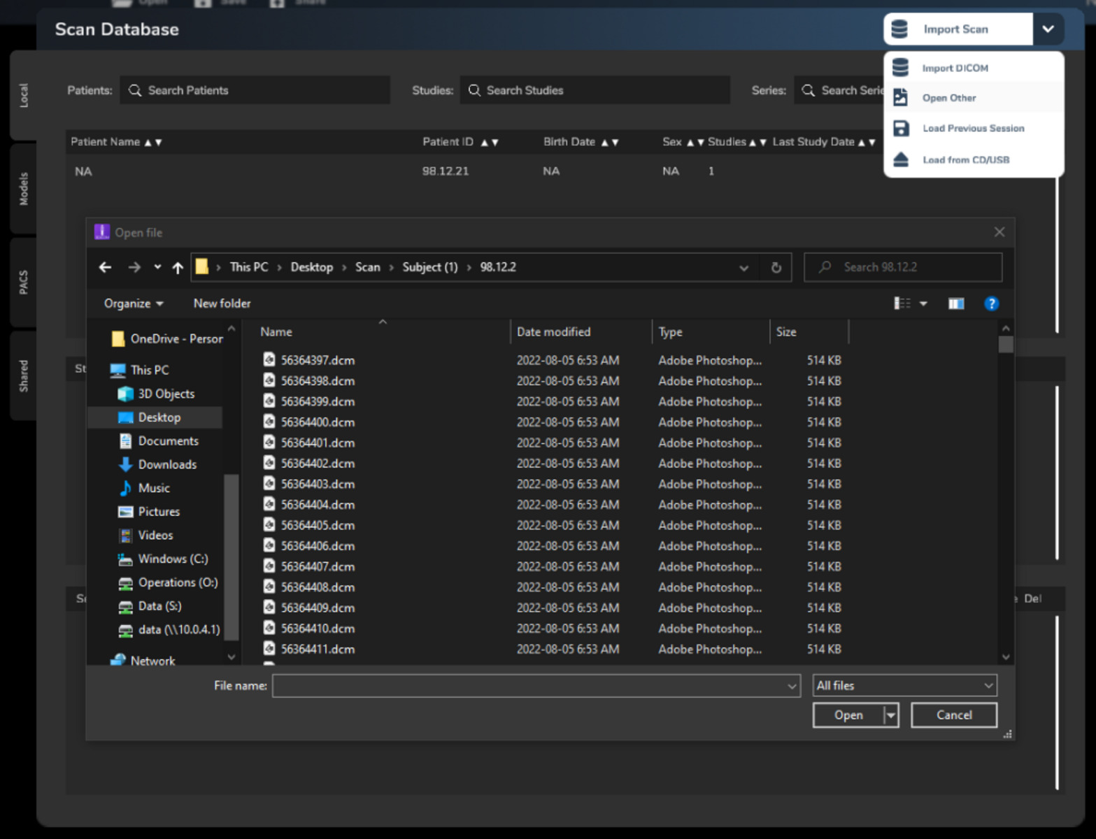
- Open the DICOM File: In the DICOM viewer software, look for an option to open a DICOM file. This is typically located in the File menu or represented by an “Open” button or icon. Click on the option and navigate to the location where your DICOM file is saved. Select the file and click “Open” to open it in the DICOM viewer.
- View and Analyze the Image: Once the DICOM file is opened, you will be able to view the medical image on your Windows computer screen. Use the tools and features provided by the DICOM viewer to enhance the image, adjust settings like brightness and contrast, zoom in or out, and analyze the image in detail. These software options often offer measurement tools, annotations, and various image manipulation functionalities.
- Additional Functions: Depending on the DICOM viewer you choose, you may have access to additional functions such as 3D reconstructions, multi-planar reconstructions, and image exporting capabilities. Explore the software’s features to make the most out of your DICOM file and its accompanying images.
Opening a DICOM file on Windows is essential for healthcare professionals, researchers, and medical students to effectively visualize and analyze medical images. The range of available DICOM viewer software ensures that users can find the application that best suits their specific requirements.
How to Open a DICOM File on Mac
If you’re using a Mac computer and need to open a DICOM file, you have several options available. Follow these step-by-step instructions to open a DICOM file on your Mac:
- Choose a DICOM Viewer: Start by selecting a DICOM viewer software that is compatible with macOS. Some popular options include OsiriX Lite, Horos, and Aeskulap DICOM Viewer. These viewers provide a user-friendly interface and a range of features for viewing and analyzing medical images in DICOM format.
- Download and Install the Viewer: Visit the website of your chosen DICOM viewer and download the installation file for macOS. Once downloaded, open the file and follow the on-screen instructions to install the viewer on your Mac. Accept any terms and conditions and choose an installation location on your computer.
- Launch the Viewer: After installation, locate the installed DICOM viewer software on your Mac and launch it. You can usually find it in the Applications folder. Double-click on the software’s icon to open it.
- Open the DICOM File: Once the DICOM viewer software is launched, look for an option to open a DICOM file. This can typically be found in the File menu or represented by an “Open” button or icon. Click on the option and navigate to the location where your DICOM file is saved. Select the file and click “Open” to open it in the DICOM viewer.
- View and Analyze the Image: Once the DICOM file is opened in the viewer, you will have access to view the medical image on your Mac screen. Use the tools and features provided by the DICOM viewer to enhance the image, adjust settings like brightness and contrast, zoom in or out, and analyze the image in detail. These software options often offer measurement tools, annotations, and various image manipulation functionalities.
- Additional Functions: Depending on the DICOM viewer you choose, you may have access to additional functions such as 3D reconstructions, multi-planar reconstructions, and image exporting capabilities. Take the time to explore the software’s features to make the most of your DICOM file and its accompanying images.
Opening a DICOM file on a Mac allows healthcare professionals, researchers, and students to efficiently visualize and analyze medical images. The availability of various DICOM viewer software options ensures that users can find the application that best meets their specific needs and preferences.
How to Open a DICOM File on Linux
Opening a DICOM file on a Linux computer is a straightforward process, as there are several DICOM viewer software options available for this operating system. Here’s a step-by-step guide to help you open a DICOM file on Linux:
- Select a DICOM Viewer: Begin by choosing a DICOM viewer software that is compatible with Linux. Some popular options include Orthanc, Horos, and Aeskulap DICOM Viewer. These viewers provide a user-friendly interface and a range of features for viewing and analyzing medical images in DICOM format.
- Install the DICOM Viewer: Depending on the DICOM viewer you choose, installation methods may vary. Some software is available directly through Linux package managers, while others may require manual installation. Refer to the documentation or website of your chosen DICOM viewer to find instructions on how to install it on your Linux system.
- Launch the Viewer: After the DICOM viewer software is installed, you can typically find it in the applications or programming section of your Linux computer. Launch the DICOM viewer by clicking on the appropriate icon or navigating to the installed directory.
- Open the DICOM File: Within the DICOM viewer, look for an option to open a DICOM file. This is usually located in the File menu or represented by an “Open” button or icon. Click on the option and navigate to the location where your DICOM file is saved. Select the file and click “Open” to open it in the DICOM viewer.
- View and Analyze the Image: Once the DICOM file is opened in the viewer, you will be able to view the medical image on your Linux screen. Use the provided tools and features to enhance the image, adjust settings like brightness and contrast, zoom in or out, and analyze the image in detail. DICOM viewer software often includes measurement tools, annotations, and image manipulation functionalities.
- Additional Functions: Depending on the DICOM viewer you choose, you may have access to advanced features such as 3D reconstructions, multi-planar reconstructions, and image exporting capabilities. Explore the software’s functionalities to utilize your DICOM file and accompanying images to their fullest extent.
By following these steps, you can open and view DICOM files on Linux effortlessly. The availability of various DICOM viewer software options ensures that Linux users can find an application that best suits their needs and preferences when working with medical images in DICOM format.
Recommended DICOM Viewer Software
When it comes to opening and viewing DICOM files, there are several DICOM viewer software options available that are highly recommended for their robust features and user-friendly interfaces. Here are a few popular DICOM viewer software programs:
- RadiAnt DICOM Viewer: RadiAnt DICOM Viewer is a powerful and feature-rich software widely used by healthcare professionals and researchers. It offers an intuitive user interface, excellent image quality, and a range of tools for viewing, analyzing, and manipulating DICOM images. RadiAnt supports advanced features like MPR (Multi-Planar Reconstruction), 3D volume rendering, and has cross-platform compatibility.
- OsiriX Lite: OsiriX Lite is a widely used open-source DICOM viewer that provides a comprehensive set of tools for primary diagnosis, research, and teaching purposes. It offers advanced visualization capabilities, such as 3D rendering, virtual endoscopy, and MIP (Maximum Intensity Projection). OsiriX Lite is well-regarded for its cross-platform compatibility and extensive plugin support.
- Horos: Horos is another open-source DICOM viewer based on the OsiriX platform. It offers a user-friendly interface, excellent performance, and advanced features for visualizing and analyzing medical images. Horos supports 3D volume rendering, MPR, and various measurement tools, making it a popular choice for medical professionals and students.
- Aeskulap DICOM Viewer: Aeskulap is a lightweight DICOM viewer with a straightforward interface and essential tools for viewing and interpreting medical images. It supports common image manipulation functions, measurement tools, and multi-frame support for animated images. Aeskulap is often praised for its simplicity and ease of use.
These recommended DICOM viewer software programs offer a range of features and functionalities to meet the diverse needs of healthcare professionals, researchers, and students. However, it’s important to note that each software has its own strengths and limitations, so it’s worthwhile to explore and compare different options to find the one that suits your specific requirements and preferences.
Alternative Methods to Open a DICOM File
While DICOM viewer software is the most common and recommended method to open DICOM files, there are alternative methods available for users who seek different approaches or face compatibility issues. Here are a few alternative methods to open a DICOM file:
- Convert to Another Image Format: If you encounter difficulties in opening a DICOM file directly, one option is to convert it to a more common image format, such as JPEG or PNG. Various online converters or image editing software can assist in this conversion process. Once converted, you can use a standard image viewer to open and view the converted file.
- Use PACS Workstations: Picture Archiving and Communication Systems (PACS) workstations are commonly used in healthcare settings for reviewing and interpreting medical images. If you have access to a PACS workstation, you may be able to import and open DICOM files directly within the workstation’s software. Consult your institution’s IT department or radiology department for guidance on accessing and using PACS workstations.
- Virtual Machine: Another option is to utilize a virtual machine (VM) that is configured with DICOM viewer software. By running a VM on your computer or through cloud services, you can create a virtual environment that supports DICOM file viewing. This method can help overcome compatibility issues or the need to install software directly on your host operating system.
- Cloud-based DICOM Viewers: There are web-based DICOM viewers available that allow you to open and view DICOM files directly from your web browser. These cloud-based viewers eliminate the need to install software on your local machine and provide the flexibility of accessing your DICOM files from anywhere with an internet connection. Some popular cloud-based DICOM viewers include Ambra Health, DICOM Grid, and MedDream Viewer.
- Open Source Libraries: For developers or individuals with programming skills, using open-source DICOM libraries can be an alternative approach to open and parse DICOM files. Libraries such as pydicom (Python), GDCM (C++), and DCMTK (C++) provide APIs that allow you to read and extract information from DICOM files programmatically.
These alternative methods can be helpful if you encounter challenges with DICOM viewer software or require different approaches for accessing and viewing DICOM files. However, it’s important to note that some methods may have limitations in terms of functionality or ease of use, so it’s advisable to assess your specific needs and consider the appropriateness of each alternative method.
Tips and Tricks for Working with DICOM Files
Working with DICOM files requires attention to detail and understanding of the specificities of this file format. Here are some tips and tricks to help you navigate and effectively work with DICOM files:
- Organize and Manage Files: Maintain a well-structured file management system for your DICOM files. Create folders to categorize images by patient, study, and series. Consistent naming conventions will simplify file retrieval and organization.
- Use DICOM Tags for Data Extraction: Take advantage of DICOM tags to extract relevant metadata from the file. DICOM tag information includes patient details, acquisition parameters, and image-specific information. Tools like pydicom in Python can assist in extracting and manipulating DICOM tags programmatically.
- Secure Patient Privacy: Ensure patient confidentiality by removing or anonymizing any identifiable information present in DICOM files before sharing or analyzing them. DICOM anonymization tools can automate this process while preserving the integrity of the image data.
- Configure Brightness and Contrast: Adjust the image brightness and contrast settings in the DICOM viewer to optimize the visualization of the medical image. Experiment with these settings to enhance image details and improve interpretation accuracy.
- Use Windowing Techniques: Employ windowing techniques to simultaneously display different components of the image, such as soft tissue and bone, to improve the visualization of specific structures or regions of interest. Window level and window width adjustments can refine the image appearance.
- Align and Compare Images: Utilize the align and compare functionalities within DICOM viewers to overlay and compare multiple images. This ability can aid in the evaluation of disease progression, treatment response, or the identification of subtle abnormalities.
- Utilize Measurement Tools: Take advantage of measurement tools provided by DICOM viewers to assess and quantify anatomical structures or lesions. These tools allow you to measure distances, angles, and areas, aiding in accurate diagnosis and treatment planning.
- Explore Advanced Visualization Techniques: DICOM viewers often offer advanced visualization techniques, such as 3D volume rendering, MPR, or maximum intensity projection. Familiarize yourself with these features to gain comprehensive insights and improve interpretation accuracy.
- Keep DICOM Viewers Up-to-date: Regularly update your DICOM viewer software to ensure compatibility with the latest DICOM standards and improve functionality. This ensures that you can take advantage of new features, bug fixes, and advancements in medical imaging technologies.
- Refer to DICOM Standards and Documentation: Familiarize yourself with the DICOM standards and documentation to understand the intricacies of the file format and its associated data elements. The official DICOM standard documentation provides detailed information on the structure and content of DICOM files.
By incorporating these tips and tricks into your workflow, you can effectively work with DICOM files, optimize image visualization, and enhance your diagnostic and research capabilities.
Potential Challenges and Troubleshooting Tips
Working with DICOM files may sometimes present challenges that can hinder your progress. Here are some potential challenges you may encounter when working with DICOM files and troubleshooting tips to help overcome them:
- Incompatibility: DICOM files can exhibit compatibility issues with certain DICOM viewer software or older systems. Ensure that you are using the latest version of the DICOM viewer software and check for updates or patches that can address compatibility concerns.
- Corrupted Files: Corrupted DICOM files can occur due to various reasons, such as transmission errors or storage issues. If you encounter a corrupted DICOM file, try to obtain an uncorrupted version from the source or consider utilizing data recovery tools specifically designed for DICOM files.
- Slow Loading or Performance Issues: Large or complex DICOM files may lead to slow loading or performance issues in DICOM viewer software. Optimize software performance by closing unnecessary applications, allocating more memory to the viewer, or utilizing software that is specifically designed to handle large DICOM datasets.
- Patient Data Privacy: Protecting patient confidentiality is paramount when working with DICOM files. Ensure that you follow privacy protocols and regulations by anonymizing or de-identifying patient data within DICOM files before sharing or storing them. Utilize DICOM anonymization tools to automate this process and maintain compliance.
- Mismatched Display Settings: DICOM files may appear differently when viewed on different displays due to variations in monitor calibration settings. To mitigate this issue, calibrate your display regularly and adjust the DICOM viewer’s display settings, such as luminance, contrast, and gamma, to achieve consistent and accurate image representation.
- Complex Data Navigation: DICOM files often contain multiple studies and series, making data navigation challenging. Utilize the DICOM viewer’s study and series sorting features to organize and streamline the data. Additionally, refer to the DICOM tags or patient information to ensure you are accessing the intended studies or series.
- Incorrect Image Orientation: DICOM files may sometimes display images with incorrect orientations. This can be resolved by using the DICOM viewer’s image rotation or flipping tools to align the image properly. Pay attention to DICOM tags that provide information about image orientation and use these as reference points.
- Limited Software Features: Some DICOM viewer software may lack certain advanced features or measurement tools required for specific use cases. In such instances, explore alternative DICOM viewer software options to find one that better suits your needs or consider utilizing additional software or plugins that complement your primary DICOM viewer.
- Data Loss or Data Management Issues: Proper data backup and management are essential when working with DICOM files. Regularly backup your DICOM files to ensure data integrity and adopt robust data management practices, such as version control and data archival, to prevent accidental data loss or corruption.
- Consult Technical Support or DICOM Experts: When facing complex or persistent issues with DICOM files, it is beneficial to consult the technical support provided by DICOM viewer software vendors or seek advice from DICOM experts. These resources can offer solutions tailored to your specific challenges.
By being aware of these potential challenges and implementing the corresponding troubleshooting tips, you can effectively navigate common hurdles and ensure a smooth workflow when working with DICOM files.